by Art Horne
Although heel cord stretching and the use of the one dimensional slant board remain mainstream in nearly every sports medicine facility and rehabilitation program following a lateral ankle sprain, the lack of ankle dorsiflexion range of motion continues to reign supreme as the underlying cause for everything from an altered gait pattern, poor squat technique and the cause of ankle reinjury itself. So if we as sports medicine and strength professionals spend so much time addressing this limitation in both our rehabilitation programs and strength routines, why then do we seem to be no further ahead when it comes to making an actual change in both true osteokinematic and arthrokinematic motion?
Well, the answer I believe lies clearly in the fact that most professionals are simply not addressing the arthrokinematic motion so closely and dearly needed within the ankle to achieve full and unrestricted ankle dorsiflexion.
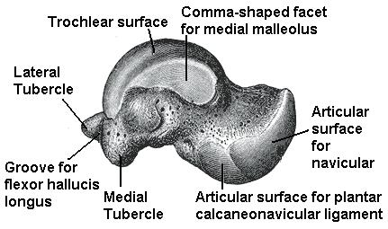
In an article by Denegar et al, the authors outline the importance of a normal joint axes of rotation along with the arthrokinematic or accessory motion around the talus itself. The authors note,
“An abnormal restrictive barrier to accessory motion changes the normal pattern of movement of a joint’s instantaneous axis of rotation. Under normal conditions, as two articulating bones glide on one another, the instantaneous axis of rotation of the joint changes accordingly. For example, as the talus glides posteriorly on the mortise, the instantaneous axis of rotation of the talocrural joint also translates posteriorly. If a restrictive barrier is encountered which limits accessory motion, the instantaneous axis of rotation becomes fixed by this restriction. Further motion thus occurs around an abnormal axis leading to subsequent joint dysfunction. At the talocrural joint, restricted posterior glide of the talus results in an abnormally anterior instantaneous axis of rotation. While full dorsiflexion range of motion may still occur, it is not necessarily reflective of normal arthrokinematic function.” (pg. 171)
So what’s next?
Well, according to the above authors, a more hands-on approach with specific attention to the talus would result in a truer movement within the talocrural joint and a much happier ankle complex.
“Our findings, and those of Green et al, have implications for rehabilitation following lateral ankle sprains as well as the risk of reinjury. All of the athletes we studied had completed a rehabilitation program as directed by their physician under the supervision of a certified athletic trainer, and had returned to sports participation. Furthermore, all had performed some form of heel-cord stretching. None, however, had received joint mobilization of the talocrural complex. Despite the return to sports and evidence of restoration in dorsiflexion range of motion, there was restriction of posterior talar mobility in most of the injured ankles. Posterior talar mobilization shortens the time required to restore dorsiflexion range and a normal gait. Without proper talar mobilization, dorsiflexion range of motion may be restored through excessive stretching of the plantar flexors, excessive motion at surrounding joints, or forced to occur through an abnormal axis of rotation at the talocrural joint.” (pg. 172)
In conclusion:
“Our results suggest that these therapeutic exercises and the passage of time restore dorsiflexion range of motion but not normal talocrural joint arthrokinematics. While more study of talocrural joint restrictions and the risk of reinjury following lateral ankle sprain is clearly needed, our results and those of Green et al suggest that (1) a restriction of talocrural arthrokinematics may be common following lateral ankle sprain; (2) the restriction my persist despite restoration of dorsiflexion range of motion; and (3) treatment of such restrictions may need to be considered in the rehabilitation following lateral ankle sprain.” (pg. 172)
The next time you encounter a patient with limited ankle dorsiflexion, whether it’s immediately after an ankle sprain or as part of your whole body assessment for another injury presentation, take a moment and assess both the quantity and quality of motion for all contributing factors to ankle motion.
Of course, if you never assess for this motion you’ll never know it’s NOT there.
Ignorance is bliss after all.
References:
Denegar, C., Hertel, J., Fonesca, J. The Effect of Lateral Ankle Sprain on Dorsiflexion Range of Motion, Posterior Talar Glide, and Joint Laxity. J Orthop Sports Phys Ther. 2002; 32(4):166-173.


