Boston Sports Medicine and Performance Group, LLC Blog

See Tomorrow's Training Technology Today

Posted by Boston Sports Medicine and Performance Group on Sat, May 5, 2012 @ 07:05 AM

Ever wonder why some Performance Coaches, Athletic Trainers, or Physical Therapists always seem to be ahead of the curve?

What do they see that you don't?

Sometimes having a little help from the sports science world and measuring progress can go a long way....

Be the first to see tomorrow's technology today at the 2012 BSMPG Summer Seminar - May 19th and 20th in Boston MA.

Click on each logo to learn more about each company and how they are changing the way we track and progress our athletes and patients.  

Stop chasing the pack, and start moving towards the front with the help from the leaders in advanced training technology from around the world!


TMG     visiblegains


ithlete          Polar


MyotestPRO          kinetic

Affectiva          BioSensics

Dartfish         zflo


Zeo            Free Lap


Tekscan      Optosource


Inside Tracker


Register for this event and for opportunities to win prizes from these sponsors today.


Seats are limited!


Click me


Topics: BSMPG, athletic training conference, boston hockey summit, Craig Liebenson, Charlie Weingroff, barefoot strength training, Andrea Hudy, Cal Dietz, Bill Knowles, Alan Grodin, Barefoot in Boston, Dan Boothby, Clare Frank

Up the Chain It Goes... (Part II)

Posted by Boston Sports Medicine and Performance Group on Fri, May 4, 2012 @ 07:05 AM

By Art Horne


turf toe derrick rose toe injury


In a follow up from a previous post (Up The Chain It Goes), additional evidence supporting the relationship within the kinetic chain has emerged from south of the equator.  In a study out of South Africa examining the link between available dorsiflexion and mechanical low back pain researchers found a statistically significant decrease in ankle dorsiflexion ROM and associated reporting of low back pain (Brantingham, 2006).   With the vast majority of adults suffering from low back pain at some time in their life, (some reports are up to 85%) and 80% of people reporting foot problems during their lifespan, it’s not a surprise to see that these two conditions may very well be related.

Let’s take a closer look:

Methods: “ The study was a blinded, 2-arm, non-randomized clinical study involving 100 subjects with chronic or recurrent mechanical low back pain (intervention group) and 104 subjects without chronic mechanical low back pain (control group) between the ages of 18 and 45.  A blind assessor performed weight-bearing goniometry of the ankle and big toe and the navicular drop test on all subjects in both groups.”

Results: “An independent t-test (inter-group) revealed a statistically significant decrease in ankle dorsiflexion range of motion in individuals with chronic mechanical low back pain.”

Conclusions: “This study’s data found that a statistically significant decrease in ankle dorsiflexion ROM, but not flatter feet, was associated with subject report of chronic mechanical low back pain disorders.”

Discussion: “The findings of this blinded study support previous reports suggesting that decreased ankle dorsiflexion may be a factor in chronic mechanical low-back pain.  There was no clear association found between decreased hallux ROM and mechanical low back pain in this study.  If these findings are confirmed through additional studies, exercise and manipulation therapy to increase ankle range of motion could become an important consideration in the treatment of some patients with mechanical low back pain disorders.”

Hmmm, if only we had some additional studies….

Perhaps this will help.

During a routine exit physical, 60 division one athletes were assessed for available weight bearing dorsiflexion bilaterally as described by Bennell et al in 1998 (inclinometer was replace by Clinometer app for ITouch) to examine limitations in this movement.   Ten athletes with limited weight bearing dorsiflexion (less than 4 inches from knee to wall) volunteered for follow up evaluation and manual treatment. Out of the initial 120 measured ankles, 47 ankles (21 right, 26 left) demonstrated limited weight bearing dorsiflexion range of motion.

Athletes were then asked to walk normally in their athletic shoes while wearing an in-shoe pressure sensor (Tekscan) and through an optical measurement system (Optojump).  Each athlete then underwent a general manual therapy intervention aimed to improve ankle dorsiflexion, followed again by the same gait analysis and pressure mapping data capture.


Gait Evaluation


Gait Cliff Notes: optimal gait should have two mountains with a trough between them. The first mountain represents heel strike to midstance, the trough representing the mid-stance phase, and the second mountain being propulsion from full foot contact to toe-off.

Easy right? Good. 

Note: The second mountain should almost always be higher than the first.


Case Study 1:

Tekscan report


Pre-treatment (RED):

Notice how the first mountain is slightly higher than the second – this is BAD!

Remember from our cliff notes: the second mountain should be higher.

Post-treatment (GREEN):

Notice change in toe off from pre- to post-treatment which specifically targeted patient's limited dorsiflexion?  The second mountain is now higher than the first. That’s a GOOD thing!

Awesome right?

Better yet – athlete was measured 3 days post treatment and improvement in Dorsiflexion range of motion stuck!  Try doing that with a slant board stretch.


See Art Horne and Dr. Pete Viteritti discuss these and other changes at the foot and ankle, and how to assess and address soft tissue and bony restrictions in their presentation at the 2012 BSMPG Summer Seminar May 19-20th in Boston.


Click me


References

Bennell KL, Talbot RC, Wajswelner H, Techovanich W, Kelly DH and Hall AJ. Intra-rater and inter-rater reliability of a weight-bearing lunge measure of ankle dorsiflexion. Australian Journal of Physiotherapy. 44;175-180.

Brantingham JW, Gilbert JL, Shaik J, Globe G. Sagittal plane blockage of the foot, ankle and hallux and foot alignment-prevalence and association with low back pain. J of Chiropractic Medicine. 2006; 4(5); 123-127.


Topics: Art Horne, basketball performance, basketball training programs, boston hockey conference, barefoot strength training, Bruce Williams, Cal Dietz, Bill Knowles, Barefoot in Boston, Chris Powers, Dorsiflexion

Up the Chain it Goes...

Posted by Boston Sports Medicine and Performance Group on Sun, Apr 29, 2012 @ 15:04 PM

By Art Horne


derrick rose torn acl


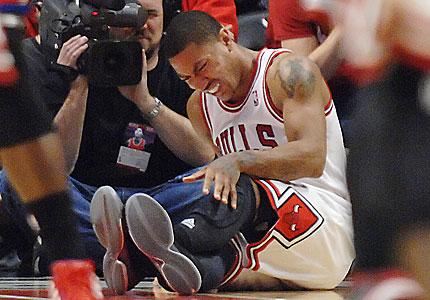
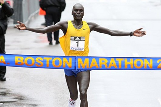
With recent season ending ACL injuries to New York Knicks Iman Shumpert, and Chicago Bull’s point guard Derrick Rose coming on the same day, (not to mention Eric Maynor from the Thunder and Spanish Star Ricky Rubio earlier this season) discussion has arisen as to how these terrible injuries could have been avoided.  Although the possible contributing factors are endless, ranging from previous injury to simply fatigue, one area worth shedding more light on, especially in the case of young Rose, is the implication of the kinetic chain as a whole.

Let’s start at the ground and work our way up.

I think we’d all agree that the big toe is a big deal.   But how closely are we looking at this “pivotal” body-ground juncture?

In a study by Munuera et al, researchers found that “Hallux interphalangeal joint dorsiflexion was greater in feet with hallux limitus than in normal feet.  There was a strong inverse correlation between first metatarsophalangeal joint dorsiflexion and hallux interphalangeal joint dorsiflexion.” (Munuera et al, 2012). 

TRANSLATION: People with abnormally stiff or limited motion at the great toe had excessive motion at the joint just distal.

If you don’t have mobility where you need it, you’ll surely get it somewhere else.

Let’s move up the chain shall we?

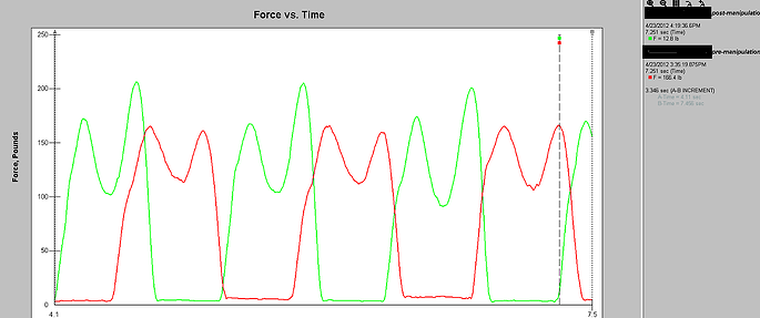
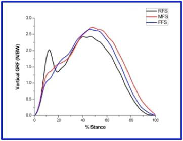
In a study by Van Gheluwe and his group,  researchers looked at how a stiff or limited great toe joint changes the way we walk.  In their study, “two populations of 19 subjects each, one with hallux limitus and the other free of functional abnormalities, were asked to walk at their preferred speed while plantar foot pressures were recorded along with three-dimensional foot kinematics.  The presence of hallux limitus, structural or functional, caused peak plantar pressure under the hallux to build up significantly more and at a faster rate than under the first metatarsal head.  Additional discriminators for hallux limitus were peak dorsiflexion of the first metatarsophalangeal joint, time to this peak value, peak pressure ratios of the first metatarsal head and the more lateral metatarsal heads, and time to maximal pressure under the fourth and fifth metatarsal heads.  Finally, in approximately 20% of the subjects, with and without hallux limitus, midtarsal pronation occurred after heel lift, validating the claim that retrograde midtarsal pronation does occur.”

TRANSLATION: if you have a limited motion in your great toe, pressure changes will occur – increase pressure changes will cause pain over time (think blister on your foot).

And pain changes the way we move – period.

Let’s take a look at the ankle.

In an article  by Denegar et al, the authors outline the importance of regaining normal talocrural joint arthrokinematics following an ankle injury.   The authors note,

 “All of the athletes we studied had completed a rehabilitation program as directed by their physician under the supervision of a certified athletic trainer, and had returned to sports participation.  Furthermore, all had performed some form of heel-cord stretching. None, however, had received joint mobilization of the talocrural complex.  Despite the return to sports and evidence of restoration in dorsiflexion range of motion, there was restriction of posterior talar mobility in most of the injured ankles.  Posterior talar mobilization shortens the time required to restore dorsiflexion range and a normal gait.  Without proper talar mobilization, dorsiflexion range of motion may be restored through excessive stretching of the plantar flexors, excessive motion at surrounding joints, or forced to occur through an abnormal axis of rotation at the talocrural joint.” (pg. 172)

TRANSLATION: I repeat, Without proper talar mobilization, dorsiflexion range of motion may be restored through excessive stretching of the plantar flexors, excessive motion at surrounding joints, or forced to occur through an abnormal axis of rotation at the talocrural joint.” (pg. 172)

If you don’t have normal ankle motion, and specifically at the talus, your ankle motion (although appearing normal) is probably coming from other joints and/or in a combination with foot pronation.


Foot Pronation = Tibial Internal Rotation

Tibial Internal Rotation = Femoral Internal Rotation

Tibia and Femur Internal Rotation  =  Knee Valgus (or knee collapse)

Knee Valgus = BAD


But just because you have some extra motion doesn’t mean you’re doomed right?

No.

But, excessive motion without the ability to control that motion certainly does.  So where does knee control come from? The Hip!

But hip strength, control, and neuromuscular timing is seldom appreciated, and in the case of the basketball athlete it is certainly poorly measured, especially after ankle injury.

In a study by Bullock-Saxton, researchers investigated muscle activation during hip extension after ankle sprain and showed a changes in timing of muscle activation in the ankle sprain grouped compared to the non-injured group.

 “the results highlight the importance of the clinician’s paying attention to function of muscles around the joints separated from the site of injury.  Significant delay of entry of the gluteus maximus muscle into the hip extension pattern is of special concern, as it has been proposed by Janda that the early activation of this muscle provides appropriate stability to the pelvis in such functional activities as gait.” (pg. 333)


In another study examining ipsilateral hip strength/weakness after the classic ankle sprain, researchers demonstrated that subjects with unilateral chronic ankle sprains had weaker hip abduction strength and less plantar flexion range of motion on the involved sides (Friel et al., 2006)

“Our findings of weaker hip abductors in the involved limb of people with chronic ankle sprains supports this view of a potential chronic loss of stability throughout the kinetic chain or compensations by the involved limb, thus contributing to repeat injury at the ankle.” (pg. 76)


“If the firing, recruitment, and strength of the hip abductor muscles in people with ankle sprains have been altered because of the distal injury, the frontal-plane stability normally supplied by this muscle is lacking, and the risk for repeat injury increases.  Weak hip abductors are unable to counteract the lateral sway, and an injury to the ankle may ensue.”

TRANSLATION: Ankle sprains cause neuromuscular changes up the chain and specifically in the hip.  If this weakness is not addressed after an ankle injury,” frontal-plane stability normally supplied by this muscle is lacking.” 


Lack of frontal-plane stability + Knee Valgus = Injury


Of course suggesting that the above points are exactly the reason for which Rose suffered his injury is certainly a stretch and not the intention of this post, nor is it to question the treatment that he or any other NBA player received prior to their devastating injury (for the record, the Chicago Bulls Sports Medicine and Strength Staff are regarded as one of the very best in the league).  What I am suggesting however is that examining athletes and patients with the use of advanced technology to determine a state of readiness to participate, and/or examine more closely changes in gait and neuromuscular firing is certainly worth pursuing, especially in light of the ever-rising salaries within professional sports.  A quick look is certainly worth the small investment.

One thing is for sure, ACL injury is not limited to teenage females or only seen on the soccer pitch.


Previous Posts:

The NBA Should Have Learned From The NFL - Injuries On The Rise

Did The NBA Lock-out Ultimately End Chauncey Billups' Career?


See lectures directly related to gait, injury prevention, and performance at the 2012 BSMPG Summer Seminar:

1. Dr. Bruce Williams: Hit the ground running: Appreciating the importance of foot strike in NBA injuries

2. Dr. Bruce Williams: Breakout Session: Restoring Gait with evidence based medicine

3. Art Horne and Dr. Pete Viteritti: Improving Health & Performance - Restoring ankle dorsiflexion utilizing a manual therapy approach

4. Dr. Tim Morgan: Biomechanics and Theories of Human Gait: Therpeutic and Training Considerations

5. Jose Fernandez: Advanced Player Monitoring for Injury Reduction


See the most advanced player monitoring equipment currently available at the 2012 BSMPG Summer Seminar:


 zeo affectiva  ithlete

BioSensics  Zflo insideTracker


Dartfish  freelap timing   Tekscanoptosource

Click me


References:

Munuera PV, Trujillo P, Guiza L, Guiza I. Hallux Interphalangeal Joint Range of Motion in Feet with and Without Limited First Metatarsophalangeal Joint Dorsiflexion. J Am Podiatr Med Assoc. 102(1): 47-53, 2012.

Denegar, C., Hertel, J., Fonesca, J.  The Effect of Lateral Ankle Sprain on Dorsiflexion Range of Motion, Posterior Talar Glide, and Joint Laxity.  J Orthop Sports Phys Ther. 2002; 32(4):166-173.


Van Gheluwe B, Dananberg HJ, Hagman F, Vanstaen K. Effects of Hallux Limitus on Plantar Foot Pressure and Foot Kinematics During Walking. J Am Podiatr Med Assoc. 96(5): 428-436, 2006.

Bullock-Saxton, J. E., Janda, V., & Bullock, M. I. (1994) The Influence of Ankle Sprain Injury on Muscle
Activation during Hip Extension. Int. J. Sports Med. Vol. 15 No. 6, 330-334.

Friel, K., McLean, N., Myers, C., & Caceres, M. (2006). Ipsilateral Hip Abductor Weakness After Inversion
Ankle Sprain. Journal of Athletic Training. Vol. 41 No.1, 74-78

Smith RW, Reischl SF. Treatment of ankle sprains in young athletes. Am J Sports Med. 1986;14:465-471.

Topics: Art Horne, basketball performance, basketball training programs, BSMPG, athletic training conference, Charlie Weingroff, boston hockey conference, barefoot strength training, Andrea Hudy, Bruce Williams, Cal Dietz, Alan Grodin, Barefoot in Boston, Dr. DiMuro, Dan Boothby, Chris Powers, achilles pain, Dorsiflexion, ankle problems

Foam Rolling and Contractile Muscle Properties by Jose Fernandez

Posted by Boston Sports Medicine and Performance Group on Wed, Apr 25, 2012 @ 06:04 AM

article by Jose Fernandez


After reading last week´s posts from Coach Boyle and Carl Valle I decided to do a little research on Self Myofascial Release and foam rolling. What motivated me is that there is not a clear protocol stablished regarding when, how and for how long athletes should use foam rollers. Some coaches recommend to roll before working out and others after, some coaches prefer to just continuously roll over the muscle surface and others recommend to hold on the trigger point for a few seconds.

What is foam rolling? (by wikipedia):

“Foam rolling is a self-myofascial release (SMR) technique that is used by athletes and physical therapists to inhibit overactive muscles. This form of stretching utilizes the concept of autogenic inhibition to improve soft tissue extensibility, thus relaxing the muscle and allowing the activation of the antagonist muscle.

It is accomplished by rolling the foam roller under each muscle group until a tender area is found, and maintaining pressure on the tender area for 30–60 seconds.”

Looking at the scientific evidence, I could not find a lot of published material either. See below some of papers I found:

A comparison of the pressure exerted on soft tissue by 2 myofascial rollers

Foam Rollers Show No Increase in the Flexibility of the Hamstring Muscle Group

The Acute Effect Of Self-Myofascial Release On Lower Extremity Plyometric Performance

In this case study we used Tensiomyography (TMG) to assess the change in contractile muscle properties (contraction time and muscle tone) after applying 2 different protocols with foam rollers. Click here to see one of my previous post with a detailed explanation about TMG and the information that it provides.

Foam Rolling & TMG Case Study:

The purpose of this case study was to analyse the change in contractile muscle properties assessed with TMG before and after applying two different SMR protocols using a foam roller. Characteristics of the roller that we used can be found here.

A professional basketball player (Age:22, H: 6.2ft, W: 198.4lbs, no injuries) with more than 6 months experience using foam rollers volunteered for the test, which was performed after a day off and consisted of assessing the Left Vastus Lateralis (dominant leg) with TMG before and after applying two different rolling interventions.

Protocol 1: Holding on the trigger point

- TMG Initial assessment on resting conditions

- Roll until the area of maximum pain is found. Hold the roll on that point for 30 seconds. Immediately after the 30 seconds continue rolling 5-6 times over the whole muscle surface

- TMG Post treatment Assessment 

Protocol 2: Cotinuous Rolling

- TMG Initial assessment

- Continuously roll over the whole muscle surface for 60 seconds

- TMG Post treatment Assessment

Restults:

Protocol 1:


Click HERE to continue reading this article...


Click me


Topics: athletic training conference, boston hockey conference, barefoot strength training, Andrea Hudy, Bruce Williams, Cal Dietz, Bill Knowles, Alan Grodin, Jose Fernandez

Heel Strikes, Sudden Impacts and Running Injuries

Posted by Boston Sports Medicine and Performance Group on Tue, Apr 17, 2012 @ 06:04 AM


If you haven't been living under a rock you know that the annual Boston Marathon took place this past Monday, April 16th.  With it came over 25 000 registered runners and thousands more non-registered runners tackling heartbreak hill and the 26.2 mile Boston course.  Although many of the leaders looked strong heading into the final mile, thousands more never experienced the thrill of victory but instead suffered the agony that so many runners face on a daily basis - a running injury that either limits or completely stops their running.

So what caused this running injury?  The answer might surprise you - it's the way you hit the ground!

Learn more about why you continue to ice your knees and shins and how you may be able to avoid these pains in the future with only a simple change in your stride.


Below is a summary of Professor Davis' article:


Do Impacts Cause Running Injuries? A Prospective Investigation

By Irene Davis, Bradley Bowser and David Mullineaux


“Up to 79% of runners sustain an injury in a given year. Approximately 80% of shod runners are rearfoot strikers. It is plausible that rearfoot strikers with increased impact loads, may be at greater risk for the development of an injury that those with lower loads. In summary, previous studies have documented higher impact loading in runner with a history of injury. However, these studies were retrospective in nature and cannot establish causative relationships. Therefore, the purpose of this prospective study was to compare the impact loads of rearfoot strike runners who go on to develop a running injury to those who have never been injured. It was hypothesized that runner who went on to develop a running- related injury would have higher vertical impact peaks, vertical average loadrates, vertical instantaneous loadrates and peak tibial shock.”

Davis and her group went on to recruit 240 female rearfoot strike runners aged 18-40 who ran a minimum of 20 miles per week.  Data was collected including ground reaction forces, vertical average loadrate, peak vertical force, etc.  Running mileage and injuries were reported monthly for 2 years.


“Our injury findings were consistent with previous literature. It has been reported that between 20-80% of runners get injured in a given year, and 57% of our runners sustained a prospective injury. In terms of injury distribution, iliotibial band syndrome, anterior knee pain, tibial stress syndrome, and plantar fasciitis were also among the top five injuries in much larger study of 2002 runners.

As hypothesized, all impact loading was greater in the injured runners compared with the never- injured group. All impact variables were significantly higher, except for VILR. Interestingly, FZ, the peak value of the vertical force, was identical between groups. This further underscores the importance of impact loads in the development of these injuries."


Barefoot in Boston

Sudden impacts are BAD! Notice the sudden spike with the RFS?

(RFS=Rearfoot Strike, FFS = Forefoot Strike, MFS = Midfoot Strike)


"The fact that all running injuries were included increases the significance of these results. While it is recognized that the etiology of running injuries is multi-factorial, this suggests that impact loading may be global indicator for the development of an injury. Based upon the odds ratio for VALR, reducing impacts is likely to result in an overall reduction of injury risk. Based on previous sports, adopting a midfoot or forefoot strike pattern will reduce these impacts. However future studies of injury patterns in midfoot and forefoot strike runners are needed.”


Want to avoid these sudden impacts associated with injury? Better think about adopting not only a different gait, but also a different pair of shoes.  Many modern running shoes have heels that are simply so thick that they prevent you from making contact with the ground in any other way than with the heel - and thus the dreaded impact forces!

Learn more about transitioning back to mother earth by reading BAREFOOT IN BOSTON: A Practical Guide to Achieving Injury Resolution and Enhancing Performance


Topics: barefoot strength training, Barefoot in Boston, barefoot running, barefoot training

Good Luck Runners! - 2012 Boston Marathon

Posted by Boston Sports Medicine and Performance Group on Mon, Apr 16, 2012 @ 07:04 AM


BSMPG wishes the over 30,000 runners in the 2012 Boston Marathon the best of luck!


Run Strong!


Boston Marathon


Topics: BSMPG, barefoot strength training, Barefoot in Boston, barefoot running, barefoot training

Christopher McDougall talks Running Barefoot and if we were BORN TO RUN

Posted by Boston Sports Medicine and Performance Group on Sun, Apr 15, 2012 @ 07:04 AM

Christopher McDougall Talks Barefoot and Why Humans Were Born to Run


Are you wondering why your running shoes resemble high heels? Ever think about why your big toe overlaps your second and why your arch really isn’t an arch anymore and resembles more of a pancake? Thinking about baring your sole? Barefoot training has recently become popularized as a potential benefit in injury prevention and rehabilitation programs. It is also purported to serve as an additional means to enhance athletic performance and running economy. However, limited clinical research is currently available to justify this practice and even less information is available describing how one may go about safely implementing a barefoot training program. This book explores the scientific and theoretical benefits concerning the merits of forgoing the modern running shoe for a simpler approach and offers real life solutions to all the obstacles standing between your feet and mother earth. Although it’s true that Americans love their shoes, what you learn about the merits of stuffing your feet and toes into these modern day casts might just have you singing a different tune – a tune your feet will certainly be much happier moving to. Welcome to Barefoot in Boston!
 
 
Enjoy Born to Run author, Christopher McDougall's TED presentation below!


Learn how you too can enjoy the benefits of being barefoot by reading BAREFOOT IN BOSTON, available now in both paperwork and kindle.

barefoot in boston

Topics: athletic training conference, athletic training, Irene Davis, Christopher McDougall, athletic training books, barefoot strength training, achilles pain, barefoot running, barefoot training

Barefoot Invades Boston!

Posted by Boston Sports Medicine and Performance Group on Fri, Apr 13, 2012 @ 19:04 PM

Boston Marathon meet Barefoot in Boston


The Barefoot Running Symposium is presented by the New England Barefoot Runners. This symposium is available to the general public at no charge on a first come, first serve basis.

This FREE event occurs during (and is part of) the 1st Annual Boston Barefoot Running Festival. This event showcases well known international speakers who have been studying running biomechanics for years. It includes pioneers of the barefoot running movement who have written books, who have previously shared their experiences through speaking engagements and barefoot running workshops.

Additional speakers are well established barefoot runners with stories of how this change in running has taken them to higher levels.

Boston Public Library
Rabb Lecture Hall
700 Boylston Street
Boston, MA 02216

Saturday, April 14, 2012
1PM – 4PM


Click HERE to read more about this exciting event and the speakers that will be there including the leader in Barefoot research - Irene Davis!

Watch Irene Davis lecture in the video below as she describes the challenges with the traditional heel strike during running and how going with less on your feet might just be the answer to your injury woes.


Not sure if baring your sole is a good idea?  Read BAREFOOT IN BOSTON, and learn how the shoes on your feet are doing more harm than good.

barefoot in boston


Topics: Art Horne, boston marathon, Irene Davis, barefoot strength training, Barefoot in Boston, achilles pain, ankle problems, barefoot training

Craig Liebenson - Keynote Speaker at 2012 BSMPG Summer Seminar

Posted by Boston Sports Medicine and Performance Group on Fri, Mar 30, 2012 @ 07:03 AM

by Craig Liebenson 


Mid-Thoracic Dysfunction: A Key Perpetuating Factor of Pain in the Locomotor System 

Dysfunction involving excessive T4-T8 kyphosis is common. Symptoms arising from regions at a distance to the mid-thoracic area are often secondary to T4-T8 dysfunction. This article will discuss why (rationale), when (indications), what (skills), and how (practical integration) T4-8 dysfunction is addressed.

Why

Mid-thoracic dysfunction involves increased kyphosis of the thoracic spine from T4-T8, usually the result of prolonged sitting in a constrained posture. Thoracic, lumbopelvic and cervicocranial posture are interrelated as links in a chain (see Figure 1). When excessive slumping becomes habitual, according to Brügger, it is called the sternosymphyseal syndrome (Lewit 1996, 1999, Liebenson et al., 1998, Liebenson 1999).


Mid-thoracic dysfunction affects the whole body's center of alignment and posture. Head and shoulder forward posture causes orofacial, neck and shoulder disorders; slumping affects breathing by leading to inhibition of the diaphragm and overactivation of the scalenes; and lumbar disc syndromes and nerve impingement have been shown to result from repetitive end-range flexion overload (Callaghan, McGill 2001).

When

Indications for treating the mid-thoracic region arise from postural analysis, passive joint mobility testing, and active joint mobility testing. The postural sign of increased thoracolumbar hypertonus is a classic sign of overactivity of the superficial "global" muscles and indicates poor "deep" muscle function (Janda 1996, Richardson 1999, Jull 2000, Hodges 2002).


Palpation of passive joint mobility and quality of end-feel is best performed in the seated position, as shown by Brügger (Brügger 2000).


The dynamic mobility screen of choice is the standing arm elevation test (Liebenson 2001).


What

Managing T4-8 dysfunction requires a broad skill set incorporating postural advice, manual manipulation, and therapeutic exercise.

Sample Exercises for Improving T4-8 Extension Mobility


  • Brügger relief position - beginner


    Brugger's Relief Position.jpg


  • Back stretch on the ball - intermediate


    T-spine Ext. on Ball.jpg
  • Kolár's wall slide with arm elevation - advanced


    Kolar's Wall Slide.jpg

    How

    Knowing why mid-thoracic dysfunction is clinically important, when it should be addressed, and what techniques are therapeutic is only the beginning point for successful management of the patient with a problem in this area. Satisfactory results will result from learning how to incorporate this knowledge and skill into patient care efficiently. A moment or two per session spent explaining the relationship between function and pain is one such step. Each exercise requires a unique "report of findings" to motivate the patient to incorporate it into his or her daily routine.

    The Brügger relief position is an ideal workplace "micro-break." It activates an entire chain of muscles linked to the upright posture. To prevent the tendency to hyperextend the lumbar spine with this exercise, it should be performed with active exhalation.

    The back stretch on the ball is comfortable and relaxing. It promotes improved respiration. It can cause dizziness at first, so the patient should be guided slowly onto it until he or she has learned how to balance on the ball.

    Kolár's wall slide with arm elevation is a functional exercise, since it combines arm elevation, squatting and breathing. Patients typically feel a nice stretch in the lattismus dorsi with this exercise.

    Summary

    T4-8 dysfunction is a common source of muscle imbalance, trigger points, joint dysfunction, and faulty movement patterns. While often asymptomatic, it is nonetheless a key source of biomechanical overload involving the neck, TMJ, shoulder, arm, and even low back regions. Treatments which aim only at the site of symptoms are bound to fail if function is disturbed due to excessive kyphosis in the mid-back.

    Rehabilitation of the upright posture is fundamental to optimization of function in the locomotor system. Neurological programs for maintenance of the upright posture are "hard-wired" into the central nervous system, making rehabilitation of the mid-thoracic area of central importance, both biomechanically and neurophysiologically. The mid-thoracic region is "linked" to a multitude of common musculoskeletal pain syndromes, and the simple assessment and treatments shown here are an excellent complement to chiropractic practice.

    References


  • Brügger A. Lehrbuch der Funktionellen Storungen des Bewegungssystems. Brugger-Verlag GmbH, Zollikon, Benglen, 2000.
  • Callaghan JP, McGill SM. Intervertebral disc herniation: studies on a porcine model exposed to highly repetitive flexion/extension motion with compressive force. Clinical Biomechanics 2001;16:28-37.
  • Hodges PW, Jull GA. Motor relearning strategies for the rehabilitation of invertebral control of the spine. In Liebenson CS. Rehabilitaiton of the Spine: A Practitioner's Manual (2nd ed). Lippincott/Williams & Wilkins, Baltimore, sched pub 2002.
  • Janda V 1996. The evaluation of muscle imbalance in Liebenson CS (ed) Rehabilitation of the Spine: A Practitioner's Manual, Lippincott/Williams and Wilkins, Baltimore, 1996.
  • Jull GA. Deep cervical flexor muscle dysfunction in whiplash. Journal of Musculoskeletal Pain 2000. 8:143-154,
  • Lewit K 1996. The role of manipulation in spinal rehabilitation in Liebenson CS (ed) Rehabilitation of the Spine: A Practitioner's Manual, Lippincott/Williams and Wilkins, Baltimore.
  • Lewit K 1999. Manipulative Therapy in Rehabilitation of the Motor System. 3rd edition. London: Butterworths.
  • Liebenson CS, DeFranca C, Lefebvre R 1998. Rehabilitation of the Spine: Functional Evaluation of the Cervical Spine, Williams & Wilkins, Baltimore.
  • Liebenson CS, Advice for the clinician and patient: The Brugger relief position. Journal of Bodywork and Movement Therapies 1999. 3:147-149.
  • Liebenson CS, Advice for the clinician and patient: Self-treatment of mid-thoracic dysfunction: a key link in the body axis. Journal of Bodywork and Movement Therapies 2001. 5:90-100.
  • Richardson C, Jull G, Hides J, Hodges P 1999. Therapeutic Exercise for Spinal Stabilization in Lower Back Pain, Churchill Livingstone. 

See Craig Liebenson speak at the 2012 BSMPG Summer Seminar this May 19-20 in Boston MA.

 Click me


Craig Liebenson  Craig Liebenson

SPONSORED BY:


zFlo


CRAIG LIEBENSON

LA Sports and Spine

Keynote Topic: Regional Interdependence: How Functional Pathology Limits Performance

Sunday Breakout Lecture: The Core as the Punctum Fixum in Sport: A Key to Making Movement Patterns More Efficient

Dr. Liebenson is an Adjunct Professor in the School of Chiropractic, Division of Health Sciences at Murdoch University, Perth Australia and consultant for the Murdoch University and the Anglo-European Chiropratic College M.Sc. program in Chiropractic Rehabilitation. The first ever chiropractic member of the McKenzie Institute (U.S.) Board of Directors, he serves on the editorial boards of numerous journals including the Journal of Occupational Rehabilitation; the PM&R  Journal of Injury, Function and Rehabilitation; the Journal of Bodywork and Movement Therapy; and Journal of Manual Therapy.

Dr. Liebenson is the first health care provider to receive a Certification of Recognition from the National Committee for Quality Assurance (NCQA) on Achievement of Recognition for Delivery of Quality Back Pain Care. He is actively engaged in ongoing research on the spinal stabilization system as a Visiting Scholar at Pr. Stuart McGill’s Spine Biomechanics Laboratory at the University of Waterloo. He regularly assists Pavel Kolar in his courses and has worked with both Dr. Karel Lewit and Pr Vladimir Janda beginning in 1987. Dr. Liebenson publishes extensively and is the editor of  the book/DVD Rehabilitation of the Spine: A Practioner's Manual (2nd ed), 2007.

He has had books published into Spanish, Greek, Korean and Japanese. He was the team chiropractor for the N.B.A. Los Angeles Clippers from the 2006-2007 season until 2009-2010 seasons and is currently a consultant for the M.L.B. Arizona Diamondbacks and Athletes Performance International.

Topics: Art Horne, BSMPG, athletic training conference, Craig Liebenson, barefoot strength training, Andrea Hudy, Bruce Williams, Alan Grodin, Dr. DiMuro, dynamic neuromuscular stabilization

Plantar Sided Foot Pain, Going Barefoot and Simple Exercises by Jeff Cubos

Posted by Boston Sports Medicine and Performance Group on Fri, Mar 16, 2012 @ 07:03 AM

I recently took part in a workshop where both manual and instrument assisted soft tissue therapy were heavily promoted as the gold standard in plantar sided foot pain presentations. While certainly such therapeutic interventions as Active Release Techniques and Graston Technique may act as positive adjuncts in the management of such cases, I often find myself using exercise interventions with relatively more rapid results. As always, patient management should always be context dependent but this is what I’m finding.

Interestingly, when therapists do consider exercise interventions, heel raises/lowers off stairs seem to be the “exercise of choice”. Often such presentations are acute or “hot” and based on Craig Purdam’s work on tendinopathies, such exercises may be too advanced for significant progress to be made.

Now before I introduce some alternative exercises, I’d like to address the “One Toe Syndrome”. Patients with plantar sided foot pain, especially women for some reason, often present with this “syndrome”. They may indeed have five toes, as most of us do, but if you look closely the toes actually function as one unit rather than five individual units.

.

The "One Toe" Syndrome

Continue to read this article by Jeff Cubos by clicking HERE.


BSMPG


Remember to Save the Date for the BSMPG 2012 Summer Seminar - May 19-20th in Boston MA.

Click me


Topics: athletic training conference, boston hockey conference, barefoot strength training, Jeff Cubos, Barefoot in Boston