Boston Sports Medicine and Performance Group, LLC Blog

Up the Chain It Goes... (Part II)

Posted by Boston Sports Medicine and Performance Group on Fri, May 4, 2012 @ 07:05 AM

By Art Horne


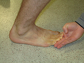
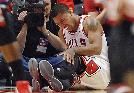
turf toe derrick rose toe injury


In a follow up from a previous post (Up The Chain It Goes), additional evidence supporting the relationship within the kinetic chain has emerged from south of the equator.  In a study out of South Africa examining the link between available dorsiflexion and mechanical low back pain researchers found a statistically significant decrease in ankle dorsiflexion ROM and associated reporting of low back pain (Brantingham, 2006).   With the vast majority of adults suffering from low back pain at some time in their life, (some reports are up to 85%) and 80% of people reporting foot problems during their lifespan, it’s not a surprise to see that these two conditions may very well be related.

Let’s take a closer look:

Methods: “ The study was a blinded, 2-arm, non-randomized clinical study involving 100 subjects with chronic or recurrent mechanical low back pain (intervention group) and 104 subjects without chronic mechanical low back pain (control group) between the ages of 18 and 45.  A blind assessor performed weight-bearing goniometry of the ankle and big toe and the navicular drop test on all subjects in both groups.”

Results: “An independent t-test (inter-group) revealed a statistically significant decrease in ankle dorsiflexion range of motion in individuals with chronic mechanical low back pain.”

Conclusions: “This study’s data found that a statistically significant decrease in ankle dorsiflexion ROM, but not flatter feet, was associated with subject report of chronic mechanical low back pain disorders.”

Discussion: “The findings of this blinded study support previous reports suggesting that decreased ankle dorsiflexion may be a factor in chronic mechanical low-back pain.  There was no clear association found between decreased hallux ROM and mechanical low back pain in this study.  If these findings are confirmed through additional studies, exercise and manipulation therapy to increase ankle range of motion could become an important consideration in the treatment of some patients with mechanical low back pain disorders.”

Hmmm, if only we had some additional studies….

Perhaps this will help.

During a routine exit physical, 60 division one athletes were assessed for available weight bearing dorsiflexion bilaterally as described by Bennell et al in 1998 (inclinometer was replace by Clinometer app for ITouch) to examine limitations in this movement.   Ten athletes with limited weight bearing dorsiflexion (less than 4 inches from knee to wall) volunteered for follow up evaluation and manual treatment. Out of the initial 120 measured ankles, 47 ankles (21 right, 26 left) demonstrated limited weight bearing dorsiflexion range of motion.

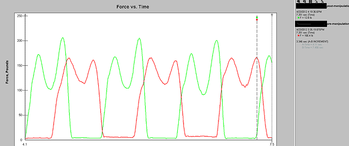
Athletes were then asked to walk normally in their athletic shoes while wearing an in-shoe pressure sensor (Tekscan) and through an optical measurement system (Optojump).  Each athlete then underwent a general manual therapy intervention aimed to improve ankle dorsiflexion, followed again by the same gait analysis and pressure mapping data capture.


Gait Evaluation


Gait Cliff Notes: optimal gait should have two mountains with a trough between them. The first mountain represents heel strike to midstance, the trough representing the mid-stance phase, and the second mountain being propulsion from full foot contact to toe-off.

Easy right? Good. 

Note: The second mountain should almost always be higher than the first.


Case Study 1:

Tekscan report


Pre-treatment (RED):

Notice how the first mountain is slightly higher than the second – this is BAD!

Remember from our cliff notes: the second mountain should be higher.

Post-treatment (GREEN):

Notice change in toe off from pre- to post-treatment which specifically targeted patient's limited dorsiflexion?  The second mountain is now higher than the first. That’s a GOOD thing!

Awesome right?

Better yet – athlete was measured 3 days post treatment and improvement in Dorsiflexion range of motion stuck!  Try doing that with a slant board stretch.


See Art Horne and Dr. Pete Viteritti discuss these and other changes at the foot and ankle, and how to assess and address soft tissue and bony restrictions in their presentation at the 2012 BSMPG Summer Seminar May 19-20th in Boston.


Click me


References

Bennell KL, Talbot RC, Wajswelner H, Techovanich W, Kelly DH and Hall AJ. Intra-rater and inter-rater reliability of a weight-bearing lunge measure of ankle dorsiflexion. Australian Journal of Physiotherapy. 44;175-180.

Brantingham JW, Gilbert JL, Shaik J, Globe G. Sagittal plane blockage of the foot, ankle and hallux and foot alignment-prevalence and association with low back pain. J of Chiropractic Medicine. 2006; 4(5); 123-127.


Topics: Art Horne, basketball performance, basketball training programs, boston hockey conference, barefoot strength training, Bruce Williams, Cal Dietz, Bill Knowles, Barefoot in Boston, Chris Powers, Dorsiflexion

Up the Chain it Goes...

Posted by Boston Sports Medicine and Performance Group on Sun, Apr 29, 2012 @ 15:04 PM

By Art Horne


derrick rose torn acl


With recent season ending ACL injuries to New York Knicks Iman Shumpert, and Chicago Bull’s point guard Derrick Rose coming on the same day, (not to mention Eric Maynor from the Thunder and Spanish Star Ricky Rubio earlier this season) discussion has arisen as to how these terrible injuries could have been avoided.  Although the possible contributing factors are endless, ranging from previous injury to simply fatigue, one area worth shedding more light on, especially in the case of young Rose, is the implication of the kinetic chain as a whole.

Let’s start at the ground and work our way up.

I think we’d all agree that the big toe is a big deal.   But how closely are we looking at this “pivotal” body-ground juncture?

In a study by Munuera et al, researchers found that “Hallux interphalangeal joint dorsiflexion was greater in feet with hallux limitus than in normal feet.  There was a strong inverse correlation between first metatarsophalangeal joint dorsiflexion and hallux interphalangeal joint dorsiflexion.” (Munuera et al, 2012). 

TRANSLATION: People with abnormally stiff or limited motion at the great toe had excessive motion at the joint just distal.

If you don’t have mobility where you need it, you’ll surely get it somewhere else.

Let’s move up the chain shall we?

In a study by Van Gheluwe and his group,  researchers looked at how a stiff or limited great toe joint changes the way we walk.  In their study, “two populations of 19 subjects each, one with hallux limitus and the other free of functional abnormalities, were asked to walk at their preferred speed while plantar foot pressures were recorded along with three-dimensional foot kinematics.  The presence of hallux limitus, structural or functional, caused peak plantar pressure under the hallux to build up significantly more and at a faster rate than under the first metatarsal head.  Additional discriminators for hallux limitus were peak dorsiflexion of the first metatarsophalangeal joint, time to this peak value, peak pressure ratios of the first metatarsal head and the more lateral metatarsal heads, and time to maximal pressure under the fourth and fifth metatarsal heads.  Finally, in approximately 20% of the subjects, with and without hallux limitus, midtarsal pronation occurred after heel lift, validating the claim that retrograde midtarsal pronation does occur.”

TRANSLATION: if you have a limited motion in your great toe, pressure changes will occur – increase pressure changes will cause pain over time (think blister on your foot).

And pain changes the way we move – period.

Let’s take a look at the ankle.

In an article  by Denegar et al, the authors outline the importance of regaining normal talocrural joint arthrokinematics following an ankle injury.   The authors note,

 “All of the athletes we studied had completed a rehabilitation program as directed by their physician under the supervision of a certified athletic trainer, and had returned to sports participation.  Furthermore, all had performed some form of heel-cord stretching. None, however, had received joint mobilization of the talocrural complex.  Despite the return to sports and evidence of restoration in dorsiflexion range of motion, there was restriction of posterior talar mobility in most of the injured ankles.  Posterior talar mobilization shortens the time required to restore dorsiflexion range and a normal gait.  Without proper talar mobilization, dorsiflexion range of motion may be restored through excessive stretching of the plantar flexors, excessive motion at surrounding joints, or forced to occur through an abnormal axis of rotation at the talocrural joint.” (pg. 172)

TRANSLATION: I repeat, Without proper talar mobilization, dorsiflexion range of motion may be restored through excessive stretching of the plantar flexors, excessive motion at surrounding joints, or forced to occur through an abnormal axis of rotation at the talocrural joint.” (pg. 172)

If you don’t have normal ankle motion, and specifically at the talus, your ankle motion (although appearing normal) is probably coming from other joints and/or in a combination with foot pronation.


Foot Pronation = Tibial Internal Rotation

Tibial Internal Rotation = Femoral Internal Rotation

Tibia and Femur Internal Rotation  =  Knee Valgus (or knee collapse)

Knee Valgus = BAD


But just because you have some extra motion doesn’t mean you’re doomed right?

No.

But, excessive motion without the ability to control that motion certainly does.  So where does knee control come from? The Hip!

But hip strength, control, and neuromuscular timing is seldom appreciated, and in the case of the basketball athlete it is certainly poorly measured, especially after ankle injury.

In a study by Bullock-Saxton, researchers investigated muscle activation during hip extension after ankle sprain and showed a changes in timing of muscle activation in the ankle sprain grouped compared to the non-injured group.

 “the results highlight the importance of the clinician’s paying attention to function of muscles around the joints separated from the site of injury.  Significant delay of entry of the gluteus maximus muscle into the hip extension pattern is of special concern, as it has been proposed by Janda that the early activation of this muscle provides appropriate stability to the pelvis in such functional activities as gait.” (pg. 333)


In another study examining ipsilateral hip strength/weakness after the classic ankle sprain, researchers demonstrated that subjects with unilateral chronic ankle sprains had weaker hip abduction strength and less plantar flexion range of motion on the involved sides (Friel et al., 2006)

“Our findings of weaker hip abductors in the involved limb of people with chronic ankle sprains supports this view of a potential chronic loss of stability throughout the kinetic chain or compensations by the involved limb, thus contributing to repeat injury at the ankle.” (pg. 76)


“If the firing, recruitment, and strength of the hip abductor muscles in people with ankle sprains have been altered because of the distal injury, the frontal-plane stability normally supplied by this muscle is lacking, and the risk for repeat injury increases.  Weak hip abductors are unable to counteract the lateral sway, and an injury to the ankle may ensue.”

TRANSLATION: Ankle sprains cause neuromuscular changes up the chain and specifically in the hip.  If this weakness is not addressed after an ankle injury,” frontal-plane stability normally supplied by this muscle is lacking.” 


Lack of frontal-plane stability + Knee Valgus = Injury


Of course suggesting that the above points are exactly the reason for which Rose suffered his injury is certainly a stretch and not the intention of this post, nor is it to question the treatment that he or any other NBA player received prior to their devastating injury (for the record, the Chicago Bulls Sports Medicine and Strength Staff are regarded as one of the very best in the league).  What I am suggesting however is that examining athletes and patients with the use of advanced technology to determine a state of readiness to participate, and/or examine more closely changes in gait and neuromuscular firing is certainly worth pursuing, especially in light of the ever-rising salaries within professional sports.  A quick look is certainly worth the small investment.

One thing is for sure, ACL injury is not limited to teenage females or only seen on the soccer pitch.


Previous Posts:

The NBA Should Have Learned From The NFL - Injuries On The Rise

Did The NBA Lock-out Ultimately End Chauncey Billups' Career?


See lectures directly related to gait, injury prevention, and performance at the 2012 BSMPG Summer Seminar:

1. Dr. Bruce Williams: Hit the ground running: Appreciating the importance of foot strike in NBA injuries

2. Dr. Bruce Williams: Breakout Session: Restoring Gait with evidence based medicine

3. Art Horne and Dr. Pete Viteritti: Improving Health & Performance - Restoring ankle dorsiflexion utilizing a manual therapy approach

4. Dr. Tim Morgan: Biomechanics and Theories of Human Gait: Therpeutic and Training Considerations

5. Jose Fernandez: Advanced Player Monitoring for Injury Reduction


See the most advanced player monitoring equipment currently available at the 2012 BSMPG Summer Seminar:


 zeo affectiva  ithlete

BioSensics  Zflo insideTracker


Dartfish  freelap timing   Tekscanoptosource

Click me


References:

Munuera PV, Trujillo P, Guiza L, Guiza I. Hallux Interphalangeal Joint Range of Motion in Feet with and Without Limited First Metatarsophalangeal Joint Dorsiflexion. J Am Podiatr Med Assoc. 102(1): 47-53, 2012.

Denegar, C., Hertel, J., Fonesca, J.  The Effect of Lateral Ankle Sprain on Dorsiflexion Range of Motion, Posterior Talar Glide, and Joint Laxity.  J Orthop Sports Phys Ther. 2002; 32(4):166-173.


Van Gheluwe B, Dananberg HJ, Hagman F, Vanstaen K. Effects of Hallux Limitus on Plantar Foot Pressure and Foot Kinematics During Walking. J Am Podiatr Med Assoc. 96(5): 428-436, 2006.

Bullock-Saxton, J. E., Janda, V., & Bullock, M. I. (1994) The Influence of Ankle Sprain Injury on Muscle
Activation during Hip Extension. Int. J. Sports Med. Vol. 15 No. 6, 330-334.

Friel, K., McLean, N., Myers, C., & Caceres, M. (2006). Ipsilateral Hip Abductor Weakness After Inversion
Ankle Sprain. Journal of Athletic Training. Vol. 41 No.1, 74-78

Smith RW, Reischl SF. Treatment of ankle sprains in young athletes. Am J Sports Med. 1986;14:465-471.

Topics: Art Horne, basketball performance, basketball training programs, BSMPG, athletic training conference, Charlie Weingroff, boston hockey conference, barefoot strength training, Andrea Hudy, Bruce Williams, Cal Dietz, Alan Grodin, Barefoot in Boston, Dr. DiMuro, Dan Boothby, Chris Powers, achilles pain, Dorsiflexion, ankle problems

Limited Anke Dorsiflexion? Find a Ninja

Posted by Boston Sports Medicine and Performance Group on Thu, Oct 6, 2011 @ 07:10 AM


By Art Horne


ankle dorsiflexion


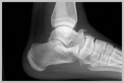
So often ankle dorsiflexion or should I say a lack thereof, is pointed at as the underlying culprit for a variety of movement impairments … and rightfully so. A lack of true talocrural motion can cause havoc up the chain involving itself in a variety of impairments including anterior tibial pain, patellofemoral pain and general low back pain to only name a few.

However, the actual limiting factor causing this lack of osteokinematic motion may be multi-factorial and if clinicians are hoping to address this limitation over the long haul with permanent change the exact location and tissue responsible for this restriction must be clearly identified and addressed with a specific intervention to match the specific tissue.

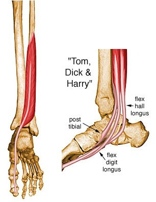
With regards to soft tissue restrictions there are only 6 possible structures that can limit this motion, and these are:

1. Soleus
2. Posterior Tibialis
3. Flexor Hallucis Longus
4. Flexor Digitorum Longus
5. Posterior Talofibular ligament
6. Posterior Tibiotalar Ligament

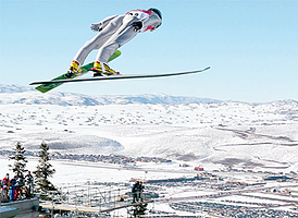
The gastrocnemieus, although probably the very first structure that comes to mind, does not limit true dorsiflexion in function (that is unless you participate in ski jumping or speed walking, and then you need to include this in your assessment), since the knee is almost always flexed when the ankle is asked to express dorsiflexion in function, such as walking, running, squatting, lunging, stepping, jumping and landing.


racewalking skijumping
 
Now that’s some dorsiflexion!


Remember, in order for your tibia to pass over your talus, and your talus to move between the tibia and the calcaneus we need to think of what pathology or dysfunction is not allowing the above mentioned tissues to lengthen.  More often than not, fibrotic adhesions within the muscles or fascial restrictions are to blame, with the filet mignon of tissue treatment choice being an Active Release Technique.  Although lesser cuts of treatment choices allow tissues to mobilize at times, rarely can a foam roller or tennis ball address a specific adhesion like a skilled clinician and the appropriate manual release technique.  That’s not to say one is wasting their time or shouldn’t employ the soft tissue mobilization techniques that they are allowed to use given their credentialing or state laws, but understanding when to refer to a specialist with a very specific skill set is the difference between a butter knife a ninja – both may get the job done but we all know which one we’d rather have on our side.


Ninja


So how does one differentiate between these tissues?

Because the Soleus and Posterior Tibialis are the two usual suspects and responsible for the majority of problems when it comes to ankle dorsiflexion limitations, these two will usually require the majority of your focus both in evaluation palpation and treatment.


posterior tibialis


However, both the Flexor Hallucis Longus and the Flexor Digitorum Longus can limit dorsiflexion and should be excluded to be sure that they are not involved.  To exclude these two structures from your list of possible dysfunctional contributors simply ask the patient to maximally dorsiflex their ankle while keeping their heel on the ground.

 
1. Gently pick up the great toe off the ground into extension. If there is slack and the patient does not indicate an increase in symptoms then the FHL is more than likely not involved.
2. Repeat tissue testing by selecting the toes and pulling them into extension.  If there is slack and the patient does not indicate and increase in symptoms then the FDL is more than likely not involved.


dorsiflexion  ankle motion


To identify the underlying tissue whether it be the soleus or posterior tibialis requires some discernable palpation skills.


Did I make a permanent change?


Charlie Weingroff calls it the “Audit Process”  while others such as good friend and colleague Pete Viteritti simply calls it, test-treat-retest.  If the correct treatment choice was matched to the correct tissue choice and location then a marked improvement in range, function and/or pain levels should occur. 

If minimal or no improvements were made than the following may have occurred:

1. You applied the correct treatment to the wrong tissue (tissue adhesion was within the posterior tibialis and you treated the soleus for example), or
2. You applied the incorrect treatment to the correct tissue (pressure was too light and thus was not sufficient to break up the underlying scar tissue), or
3. The limiting factor was not soft tissue but instead an osteokinematic “misalignment” or a position fault as described and made famous by Brian Mulligan (more Mulligan in a future post).

Summary: The most important step in any treatment approach starts with first identifying the correct pain generator or dysfunctional tissue involved.  Without a correct place to start, all treatment options will fail to make a lasting change.

Topics: Art Horne, basketball conference, basketball training programs, athletic training conference, athletic training, boston hockey conference, Barefoot in Boston, Dorsiflexion, ankle problems