Boston Sports Medicine and Performance Group, LLC Blog

Essential Reading Spotlight - Greenman's Principles of Manual Medicine

Posted by Boston Sports Medicine and Performance Group on Wed, Oct 12, 2011 @ 06:10 AM

I have to admit I missed the boat on the importance of manual medicine for a long time.  After speaking to the best clinicians and therapists in the area there was one book that each and everyone of them, regardless of their current treatment approach, recommended I read - Greenman's Principles of Manual Medicine (Point (Lippincott Williams & Wilkins))   

After recently completing this book I now know why.  This book is a must for anyone treating musculoskeletal dysfunction on a daily basis.  Below is a short excerpt from this book.


The Manipulable Lesion

"The acceptable term for this entity is somatic dysfunction. It is defined as impaired or altered function or related components of the somatic (body framework) system; skeletal, arthrodial, and myofascial structures; and related vascular, lymphatic, and neutral elements. Notice that the emphasis is on altered function of the musculoskeletal system and not on a disease state or pain syndrome." pg 11


DIAGNOSTIC TRIAD FOR SOMATIC DYSFUNCTION 


"The mnemonic ART can express the diagnostic criteria for identification for somatic dysfunction.

“A” stands for asymmetry of related of the musculoskeletal system, either structural or functional.
“R” stands for range of motion of a joint, several joints, or region of the musculoskeletal system. The range of motion could be abnormal by being either increased (hypermobility) or restricted (hypomobility). The usual finding in somatic dysfunction is restricted mobility, identified by observation and palpation using both active and passive patient cooperation.
“T” stands for tissue texture abnormality of the soft tissues of the musculoskeletal system (skin, fascia, muscle, ligament, etc.). Tissue texture abnormalities are identified by observation and a number of different palpatory tests.

Some authors add one of two other letters to this mnemonic. “P” or a second “T”. “P” stands for pain associated with other findings, and “T” stands for tenderness on palpation of the area. Tenderness is particularly diagnostic if localized to a ligament. A normal ligament is not tender. A tender ligament is always abnormal. However, both pain and tenderness are subjective findings instead of the objective findings of symmetry, altered range of motion, and tissue texture abnormality. By the use of these criteria, one attempts to identify the presence of somatic dysfunctions, their location, whether they are acute or chronic, and particularly whether they are significant for the state of the patients wellness of illness at that moment in time. In addition to the diagnostic value, changes in these criteria can be of prognostic value in monitoring the response of the patient, not only to manipulative treatment directed toward the somatic dysfunction, but also to other therapeutic interventions."  Pg 11-12


Topics: Art Horne, basketball performance, basketball resources, BSMPG, athletic training conference, boston hockey summit, boston hockey conference, barefoot strength training, barefoot training

Interview with Mark Toomey from Dragondoor.com

Posted by Boston Sports Medicine and Performance Group on Mon, Oct 10, 2011 @ 06:10 AM


Mark Toomey


This past June Mark Toomey and Dr. John DiMuro presented at the BSMPG summer seminar, "Standing On The Shoulders Of Giants."  Below is a small portion of Mark's most recent interview.


"We presented a paper together at Boston University and Northeastern University in June on fostering collaborative efforts between medical and exercise professionals. We "knuckle draggers" and I'm proud to call myself that, don’t have to play doctor. There are medical professionals who desire a relationship with us as long as we let them know what we're good at. My swim lane is only this wide but it’s this deep. I don’t want to be a doctor, I don’t want to be a healer - that’s a physician's job. But there is a legitimacy that creating a relationship with the medical community can give us."

Continue to read this interview on Dragondoor.com by clicking HERE

Topics: Art Horne, athletic training conference, athletic training, athletic training books, Mark Toomey, Barefoot in Boston, Dr. DiMuro

Help Yourself.... So You Can Continue To Help Others

Posted by Boston Sports Medicine and Performance Group on Sat, Oct 8, 2011 @ 07:10 AM


By Art Horne


athletic training


We’ve all done it from time to time – missed lunch to work with an athlete, stayed up late to write notes on the previous day’s work or ignored a pending physician or dentist appointment because we just didn’t have time.

And although it’s usually celebrated by those you care for and rewarded by the boss everyone knows this downward cycle of ignoring one’s own health in favoring of attending to the health of others can only be headed in one direction. 

So the next time you board a plane when traveling remember to listen to your flight attendant’s very important message,

“During a change in altitude, oxygen masks will drop down from the ceiling.  For patients travelling with young children, be sure to put your mask on first before helping your child.”


As busy as we all are it’s important to remember that the health of those we provide services for is inherently tied to our very own.  It’s tough to continue to help others if you haven’t first helped yourself. 

Topics: Art Horne, basketball conference, BSMPG, athletic training conference, athletic training books, barefoot strength training

Limited Anke Dorsiflexion? Find a Ninja

Posted by Boston Sports Medicine and Performance Group on Thu, Oct 6, 2011 @ 07:10 AM


By Art Horne


ankle dorsiflexion


So often ankle dorsiflexion or should I say a lack thereof, is pointed at as the underlying culprit for a variety of movement impairments … and rightfully so. A lack of true talocrural motion can cause havoc up the chain involving itself in a variety of impairments including anterior tibial pain, patellofemoral pain and general low back pain to only name a few.

However, the actual limiting factor causing this lack of osteokinematic motion may be multi-factorial and if clinicians are hoping to address this limitation over the long haul with permanent change the exact location and tissue responsible for this restriction must be clearly identified and addressed with a specific intervention to match the specific tissue.

With regards to soft tissue restrictions there are only 6 possible structures that can limit this motion, and these are:

1. Soleus
2. Posterior Tibialis
3. Flexor Hallucis Longus
4. Flexor Digitorum Longus
5. Posterior Talofibular ligament
6. Posterior Tibiotalar Ligament

The gastrocnemieus, although probably the very first structure that comes to mind, does not limit true dorsiflexion in function (that is unless you participate in ski jumping or speed walking, and then you need to include this in your assessment), since the knee is almost always flexed when the ankle is asked to express dorsiflexion in function, such as walking, running, squatting, lunging, stepping, jumping and landing.


racewalking skijumping
 
Now that’s some dorsiflexion!


Remember, in order for your tibia to pass over your talus, and your talus to move between the tibia and the calcaneus we need to think of what pathology or dysfunction is not allowing the above mentioned tissues to lengthen.  More often than not, fibrotic adhesions within the muscles or fascial restrictions are to blame, with the filet mignon of tissue treatment choice being an Active Release Technique.  Although lesser cuts of treatment choices allow tissues to mobilize at times, rarely can a foam roller or tennis ball address a specific adhesion like a skilled clinician and the appropriate manual release technique.  That’s not to say one is wasting their time or shouldn’t employ the soft tissue mobilization techniques that they are allowed to use given their credentialing or state laws, but understanding when to refer to a specialist with a very specific skill set is the difference between a butter knife a ninja – both may get the job done but we all know which one we’d rather have on our side.


Ninja


So how does one differentiate between these tissues?

Because the Soleus and Posterior Tibialis are the two usual suspects and responsible for the majority of problems when it comes to ankle dorsiflexion limitations, these two will usually require the majority of your focus both in evaluation palpation and treatment.


posterior tibialis


However, both the Flexor Hallucis Longus and the Flexor Digitorum Longus can limit dorsiflexion and should be excluded to be sure that they are not involved.  To exclude these two structures from your list of possible dysfunctional contributors simply ask the patient to maximally dorsiflex their ankle while keeping their heel on the ground.

 
1. Gently pick up the great toe off the ground into extension. If there is slack and the patient does not indicate an increase in symptoms then the FHL is more than likely not involved.
2. Repeat tissue testing by selecting the toes and pulling them into extension.  If there is slack and the patient does not indicate and increase in symptoms then the FDL is more than likely not involved.


dorsiflexion  ankle motion


To identify the underlying tissue whether it be the soleus or posterior tibialis requires some discernable palpation skills.


Did I make a permanent change?


Charlie Weingroff calls it the “Audit Process”  while others such as good friend and colleague Pete Viteritti simply calls it, test-treat-retest.  If the correct treatment choice was matched to the correct tissue choice and location then a marked improvement in range, function and/or pain levels should occur. 

If minimal or no improvements were made than the following may have occurred:

1. You applied the correct treatment to the wrong tissue (tissue adhesion was within the posterior tibialis and you treated the soleus for example), or
2. You applied the incorrect treatment to the correct tissue (pressure was too light and thus was not sufficient to break up the underlying scar tissue), or
3. The limiting factor was not soft tissue but instead an osteokinematic “misalignment” or a position fault as described and made famous by Brian Mulligan (more Mulligan in a future post).

Summary: The most important step in any treatment approach starts with first identifying the correct pain generator or dysfunctional tissue involved.  Without a correct place to start, all treatment options will fail to make a lasting change.

Topics: Art Horne, basketball conference, basketball training programs, athletic training conference, athletic training, boston hockey conference, Barefoot in Boston, Dorsiflexion, ankle problems

Standing On The Shoulders Of Giants DVD's Available

Posted by Boston Sports Medicine and Performance Group on Thu, Sep 29, 2011 @ 08:09 AM

Missed this summer's BSMPG summer seminar?

Wished you could have seen the authors of these books and video speak while they were in Boston?


Tom Myers  Charlie Weingroff  Clare Frank

Well now you can - We captured three groundbreaking presentations from this once in a lifetime seminar.  Catch three of these Giants in action as they presented at the 2011 "Standing On The Shoulders Of Giants" Summer Seminar.

This conference was held in Boston, MA on June 3rd and 4th, 2011

Included: 3 DVDs with over 4.5 hours of Sports Medicine and Rehabilitation Information

Presentations by: Clare Frank - Muscle Balance and Stability, Tom Myers - Anatomy Trains and Myofascial Fitness and Charlie Weingroff - Trying to Define the Core.

clare frank Tom Myers Charlie Weingroff

Click HERE for complete details.

Topics: Art Horne, basketball resources, basketball conference, BSMPG, boston hockey summit, athletic training, Charlie Weingroff, boston hockey conference, Tom Myers, athletic training books, barefoot strength training, Clare Frank

SAVE THE DATE : March 30th - April 1st, 2012

Posted by Boston Sports Medicine and Performance Group on Wed, Sep 28, 2011 @ 09:09 AM

BSMPG


BSMPG is busy working on the final details of a continuing educational opportunity that you won't want to miss!

Pencil in this 3-day learning event coming March 30th through April 1st of next year. 

This event will only be open to 30 individuals and 30 individuals only.

NO walk-ins. NO casual learners. NO nodding off after lunch is served. 

This will be an intense 3-day course developed for those skilled clinicians looking to take their treatment approach to the next level and is the first of it's kind on the east coast!

Location: Northeastern University, Boston MA, USA

Details coming soon!

Topics: Art Horne, BSMPG, athletic training conference, Charlie Weingroff, Clare Frank

Integrated Care - Part VI: Getting Everyone To Squat

Posted by Boston Sports Medicine and Performance Group on Tue, Sep 27, 2011 @ 07:09 AM


by Art Horne

Since squatting and the decision to squat or not comes up more often than any other subject when it comes to sports medicine and performance professionals, we decided to tackle this subject head on in this latest post.  Everyone should be able to squat - there I said it. So whether you'll be teaching it after surgery, or training it in the weight room it's important that the beliefs and concepts that you have pertaining to the squat is the same as those that you work with in order to achieve the highest level of athlete success. 

Below is an outline which we have used in a staff journal club to finalize our teaching progressions and teaching cues for the squat pattern while also getting everyone on the same page and on board with the importance of squatting.

sports medicine


Squat Goal: all athletes should be constantly working towards a score of three on their OH Squat test. Whether they initially present to Sports Medicine with back, knee, hip or other LE injury or pain, all care and performance training providers should be able to look at this movement pattern and address any movement concerns IN ADDITION to their traditional rehab or performance program regardless of initial pain or injury presentation.

FMS Squat Test:  see functionalmovement.com  for complete details
(Have a staff member certified or familiar with the FMS review testing criteria, followed by staff members from both staff’s testing and evaluating each other).


What if the student-athlete describes PAIN during the squat test?


sports medicine


Only qualified medical professionals should deal with pain and painful movement patterns.  If a student-athlete experiences pain they should be referred to the athletic trainer coordinating their care at the college level or another health care professional if you are working in the private sector.  Although many “painful” maneuvers are often neglected and played down by the student-athlete, seldom do those with pain during a simple bodyweight squat ever go on to compete at a high level without future injury and time loss.

Since PAIN is complicated, multifactorial and often unpredictable, a painful pattern should be “broken down” to its component parts so to find the underlying pain generator or limiting factor.  The Selective Functional Movement Assessment TM offers health care providers a systematic approach and a common movement language among all staff members.

(Have a staff member certified or familiar with the SFMA squat breakout present this algorithm to the remainder of your staff and then follow up by having individuals from both groups evaluate each other while identifying the limiting factor within each individual’s squat pattern.


SFMA Squat Breakout:


1. Overhead deep squatting pattern

2. Fingers interlocked behind neck into deep squat position
a. This position lowers the difficulty by not having the shoulders flexed vertical
b. Removes the upper body components and reduces the level of dynamic stability needed to perform the squat.

3. Assisted Squat from ATC into deep squat
a. Athlete holds hands of evaluator while performing squat.
b. Looks at the true symmetrical mobility of the lower body (hips, knees, ankles) without the requirement of dynamic stability.

4. Half kneeling Dorsi-flexion (test for ankle ROM)
a. Place foot on weight bench and lean forward over the foot as far as possible without the heel coming off the bench.
b. The knee should move forward out past the toes at around 4 inches.
c. Repeat for both sides.

5. Supine Knees-to-chest – test for anatomical limitations at hip in non-weight bearing position.
a. Looks at mobility of the hips, knees and spine in a non-weight bearing position.
b. Can help differentiate a mobility vs. stability problem
c. Have the patient grab both shins and attempt to touch their thighs to their lower rib cage.
d. If they can’t get their calves to touch their thighs due to knee tightness, have them grab their thigh instead and repeat thin maneuver. (This will help differentiate between hip and knee mobility issues. If in this position they still can’t get their thighs to touch their rib cage, then there is a potential hip dysfunction).


What’s Next?

Since so many of us sit on our butts all day it’s no surprise that our butts are rarely working to the level they should be. 

(Unless of course you use your butt for a simple paper weight – then your butt works just fine).

Because the squat is the end goal for all athletes in terms of both health and performance, it’s important for athletic trainers and other health care professionals to begin grooving this pattern early on in the rehabilitation process so that there is not only a seamless transition from one Sports Medicine to Performance Training, but also a clear message when it comes to cueing and technique among all professionals on staff.  In order to have everyone speaking the same squat language, starting everyone back on the ground and building a solid and logical progression from post-surgical all the way to high performance only makes sense while the entire staff is playing nice together.

Below is a starting point with a few teaching cues to get the conversation started and a solid progression built between the two groups.   (I have purposefully not included a complete progression below so to force the issue with your own staff.  People tend to complete tasks fully if they have had an opportunity to contribute to so be sure to ask for feedback from both staffs – Sports Medicine and Strength & Conditioning).


Back to Basics: (Supine Table Series)


1. Teach Glute Max w/ Bridge
2. Supine Bridge w/ Thera-band
3. Supine Bridge w/ Marching
4. Single Leg Hip Bridge


Teaching Points:


a. Squeeze Glutes to hold “gold coin” between butt cheeks or pretend to “crack a walnut” while bridging
b. brace core musculature as taught by Stu McGill
c. Ensure hamstrings are not dominate during exercise by palpating medial tendons.
d. Add a theraband around knees or create an ADDuction force to the knees to facilitate additional glute max contraction


– NEVER PLACE A BALL BETWEEN THE KNEES!


Teaching Progression for the Troubled Squat


gobletsquat


1. Goblet Squat: hold Kettlebell by the horns and squat to depth (providing a weight to the anterior portion of body balances the athlete and gives an additional point of stability

2. Descend squat so that elbows touch VMO of each knee.

3. If depth is a problem “wedge” elbows inside of knees to mobilize the hips.

4. In the early stages be sure to stand behind your athlete to ensure safety – encourage thoracic extension and a flat back

5. ** use clinical judgment based on patient presentation and injury. If unsure, REGRESS activity and PERFECT – OWN THE MOVEMENT before progressing to more advanced alternatives.


READ:  Squatting - An Expression Of Health


Next week: Bringing both staffs together to learn and master the hip hinge – a must for avoiding and rehabilitating back pain and also pulling big deadlift numbers!


Topics: Basketball Related, Art Horne, basketball resources, BSMPG, athletic training conference, athletic training, FMS, Barefoot in Boston

Integrated Care - Part V: When NOT Touching Your Toes is NOT a Hamstring Problem

Posted by Boston Sports Medicine and Performance Group on Sun, Sep 18, 2011 @ 06:09 AM

by Art Horne


I remember being taught in school that people have low back pain because their hamstrings are tight.  Therego, stretch the hamstrings and resolve the low back pain.  It was a simple solution to a very complex and often misunderstood problem and yet as a student it was a clean and direct resolution to an often nagging dilemma.

Further testing and evaluation always “proved” the hamstrings as the culprit since so many of the low back pain patients were never able to touch their toes, and of course toe touching was a direct result of hamstring length.  Again, this bore out to make sense since after spending an exhausting amount of time stretching these patient’s hamstrings, and then retesting, some measurable change were noted (sometimes) with the patient inching closer to their toes.  The only problem in this clean and concise “hamstring-LBP” relationship was that these patients, no matter how long I stretched them or which stretching technique I employed ALWAYS ended up coming back the very next day for the same stretching routine and the same unresolved back pain.


BSMPG

Insanity: Doing the same thing over and over again and expecting different results.


Of course, many of these patients really never had “tight” hamstrings after all, but instead had hamstrings that were preventing them from a place they no had no business being in the first place. 

What do I mean?

Well, many of these patients, or at least the majority of them,  were flexion intolerant which means both flexing their spine and/or moving into flexion caused a reflexive “tightening” of the hamstrings to essentially keep them from FLEXING FORWARD and moving into a region which would exacerbate their current condition, or “a place they have no business being.”  In addition, many of these “tight hamstring” patients were never ever feeling a hamstring stretch, but instead were experiencing a neural stretch, which unfortunately after stretching only continued their “tight-stretch-pain-tight” cycle. 

Soo… Should I Stretch or Not?

1. First distinguish between hamstring tightness and neural tension.  Neural tension is always described differently than muscular tightness – that is if you’re listening.  I had a professor who told me that your patient will always tell exactly what is wrong with them if you listen long enough.  And if you listen just a little bit longer, they’ll also tell you exactly how to make them better.  This case is no different – of course, if you’re too busy catering water you’ll never have enough time to listen long enough.

2. Neural tension is often described as “pinching” or with other words that clearly denote nerve origin such as “zapping” or “burning.”  Muscle tension doesn’t zap or burn.

3. Confirm neural involvement with a slump test – then STOP stretching and begin a nerve flossing regiment if indicated.

4. Does the patient need more mobility (hamstring stretching) or do they simply lack the appropriate stability (or neuromuscular control) which ultimately is limiting them from moving into a place where they simply didn’t have the requisite control? I’m willing to bet that more often than not that stability/neuromuscular control is the limiting factor and not hamstring length – especially in low back pain patients.

So what does this have to do with integrated care?

1. On evaluation during pre-participation screenings a simply toe touch as a gross indicator of both ability and willingness to forward flex will “catch” those athletes who either have had previous back pain, current back pain or a complete lack of understanding relating to lumbar spine/pelvis position and an ability to disassociate them during normal daily living.

2. Those with pain or previous pain should be referred to sports medicine for further evaluation – yes, even those that don’t currently have pain.

3. Those that have trouble separating hip and lumbar motion should be placed into a “teaching” group while in the weight room until they can successfully “stiffen” their spine while gaining mobility and motion throughout the hips; both which are essential if any type of squat or deadlift pattern are prescribed in their program.  But really more importantly, these two patterns occur each and every day of their lives and must be grooved before major lumbar spine pathology presents itself.

Click HERE for a quick note on establishing a hip hinge.


Next week: A quick in-service to get both Sports Medicine and Strength Training hinging at the hips and teaching all athletes a great squat pattern. 

Topics: Art Horne, basketball performance, basketball resources, basketball conference, basketball training programs, athletic training conference, barefoot strength training, Barefoot in Boston, barefoot training

Integrated Care - Part IV : Addressing & Correcting Hip Extension Problems

Posted by Boston Sports Medicine and Performance Group on Mon, Sep 12, 2011 @ 07:09 AM

by Art Horne


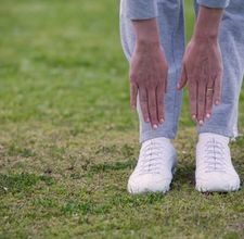
As we mentioned last week, our society has clearly become hip flexion dominant. 

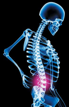
This is really no surprised as Janda identified this “epidemic” long ago and termed it, the Lower Crossed Syndrome.  Clearly ahead of his time, and well before Blackberrys and IPhones caused us to hunch over and run into people on the sidewalk, Janda also described and discussed the upper crossed syndrome which is more prevalent today than ever as well.  With that said, so many of the young “healthy” athletes that sign up to play collegiate level athletics no longer are able to express the fundamental movement patterns that we so often take for granted.  This of course is not always a mobility problem, as many athletes are not able to reach end range of these patterns simply out of a reflexive protective mechanism.


sports medicine resources


Your body will simply not allow you to go where you have no business going.  (Ever wonder why LBP patients can’t touch their toes? Hint: it has nothing to do with hamstring length and everything with your brain not letting you get to end range flexion, you know, the place you have no business going)
In other cases, mobility is the main culprit and can usually be addressed with a simple movement exam along with some corrective therapy and exercises. 


Let’s take a look at an example to see what I mean.

Case Presentation:

This athlete presented to me many years ago, and unfortunately the overall theme continues year after year despite our best efforts to educate our athletes and their high school and youth coaches.

Here’s the story:

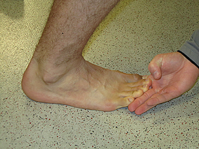
On evaluation athlete complains of having a persistent anterior hip pain from day one of pre-season practice.  She states that she had a “significant” hip injury at age 13 which lasted about one year and limited her from all sporting activities including gymnastics where she originally hurt herself during a coach “assisted” stretch.  At the time of the stretch, the athlete’s injured leg was down and extended behind her pelvis, with knee at 90 degrees and the opposite limb forced into extreme flexion.  At that time she felt intense pain and was not able to return to any physical activities for about one year. 

She went on to a successful high school career and eventually earned a college scholarship for her efforts.


sports medicine

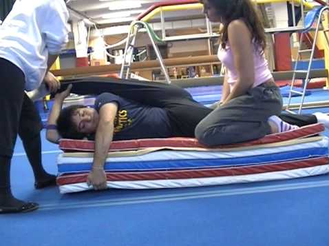
(not the same stretch - but close. OUCH!) 


movement screen


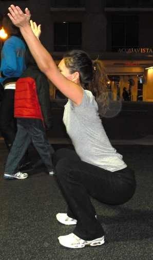
On movement evaluation utilizing the SFMA, cervical and shoulder motions were FN (functional and non-painful), multi-segmental flexion FN, multi-segmental extension FP (functional but painful), multi-segmental rotation DN (dysfunction and non-painful) away from the injured side, single leg stance was DP on injured side, FN on uninjured side (NOTE: during this test athlete complained of pain with standing hip flexion.  She was however able to get her knee/femur past 90 degrees of hip flexion but had to first abduct her leg then lift it in front – so to basically avoid iliacus involvement and use only psoas with some help from TFL and Sartorius). Lastly,  deep squat was DN. 


(Now, according to the SFMA I should have “broke out” her multi-segmental rotation pattern and addressed her subsequent restriction but given her prior history and description of pain I decided to go directly to the prone hip extension test to confirm my suspicion that she had originally injured her iliacus some 5 years ago.)

On prone hip extension, athlete was unable to extend her injured leg to a minimum of 10 degrees.

Treatment Plan: evaluate and address tissue density changes and restrictions within the hip flexion musculature including both psoas and iliacus specifically.


If you aren’t familiar with manual therapy techniques to address soft tissue restriction within the iliacus consult a co-worker or expert in your area for help or training (If you’re in the Boston area one of the most talent manual therapist I’ve ever had the pleasure working with is Dr. Pete Viteritti.


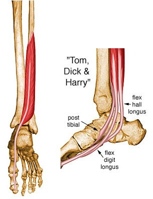
Below are a few key technical points regarding treatment of the Iliacus utilizing a manual therapy release technique.  Remember: the iliacus is to hip dysfunction as the psoas is to lumbar dysfunction.

sports medicine


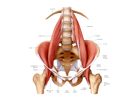
1. Begin with the patient lying on their side, hip and knee flexed and relaxed.

2. With the contact fingers extended, work the soft contact from the anterior superior iliac spine (above the inguinal ligament) medically onto the iliacus treating from proximal all the way distal to the lesser trochanter.  The adhesion can be anywhere in the muscle.  Also, be sure to move your hand contact treating medially until you bump into the psoas.  The junction of the iliacus and psoas is very important, be sure they are not adhering to one another. (adhesion's between muscles which cause them to adhere to one another is much more of a problem than an adhesion in a muscle itself).

3.The inguinal ligament should also be checked to be sure you can bow it both distal and proximal, as it can adhere to the iliacus underneath it.  Find the inguinal ligament at the ASIS and trace it as it moves medially and deep.  It is only the lateral aspect of it that comes in contact with the iliacus and can become entrapped.

4. As you begin, be sure to move the mesentary medially and not treat through it.  Use care to avoid putting tension on the mesentary as this will not only cause discomfort to the patient, but will significantly limit treatment effectiveness.

5. Once on the tissue, begin to put tension on the tissue superiorly with your inferior hand while the superior hand backs it up.

6. Have the patient extend the hip and knee straight and then extend the hip as far as possible.

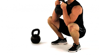
Post treatment: Athlete was able to regain full prone hip extension, pain resolved with both single leg stance (athlete was able to lift knee/leg straight up in sagittal plane) and multi-segmental extension pattern.  Deep squat pattern improved significantly but was not yet perfect.  And most impressive post treatment was the look of shock and excitement on her face.

Whether you’re dealing with a shortened iliacus, a tight psoas major or a restricted rectus femoris (or perhaps even a shortened rectus abdominis thanks to the 2 million crunches you’d done), identifying the global limitation first (an extension pattern in this case), and then referring to an expert or addressing the underlying tissue restricting this pattern yourself before high levels of organized activity begins can mean the difference between weeks of treatment post injury or a few moments of your time prior, during your screening process.  Of course identifying the exact limiting factor/tissue/pain generator is the ultimate factor when it comes to whether your treatment will be a success or not.

“So what does this have to do with integrated care?  This sounds like a pure sports medicine problem and treatment approach to me.”

Perhaps – but all strength coaches can look at global movement patterns including extension and make the appropriate referrals.  Whether it’s during your pre-participation examination or during a simple recheck in the weight room – having all coaches, athletic trainers and therapists understanding the normal parameters of human movement and speaking the same language eliminates the language barrier and allows all parties involved in the care and performance of the student-athlete to be provide a unified care approach to the identified problem.  Although many strength coaches won’t be able to apply a manual therapy technique for this identified problem, appropriate strategies within the weight room can certainly maintain this new tissue quality and “cement” this new found range of motion with strength exercises appropriate for the athlete and previous injury.

Although the skill set or specific treatment modality between the two professional groups my vary slightly, the underlying philosophy should not and in this case addressing this extension limitation with whatever tools you are allowed to use will certainly pay dividends at the end of the day.


Next Week: When Not Being Able To Touch Your Toes Is Not A Hamstring Issue


Topics: Art Horne, basketball training programs, athletic training conference, basketball videos, Pete Viteritti, athletic training books, barefoot strength training, barefoot running

Art Horne Interviews with Joe Heiler on SportsRehabExpert.com

Posted by Boston Sports Medicine and Performance Group on Wed, Sep 7, 2011 @ 07:09 AM


sportsrehabexpert


Click HERE to listen to this interview.

In this interview, Art discusses his new book, "Barefoot in Boston: A Practical Guide to Achieving Injury Resolution and Enhancing Performance", as well as discussing some research on barefoot walking and training versus wearing shoes, what to look for in minimalist shoes, how he has incorporated barefoot training with his basketball team, and much more..(including how to take a hack saw to a basketball shoe!)

Topics: Basketball Related, Art Horne, BSMPG, functional movement screen, boston marathon, foot pain, foot fracture, barefoot strength training, Barefoot in Boston, achilles pain, barefoot running, barefoot training