Boston Sports Medicine and Performance Group, LLC Blog

Did The NBA Lock-out Ultimately End Chauncey Billups' Career?

Posted by Boston Sports Medicine and Performance Group on Tue, Feb 7, 2012 @ 21:02 PM


By Art Horne


chauncey billups


It’s official.

The NBA has lost another of its premiere players to injury.

Chauncey Billups has suffered a season-ending, and most likely a career-ending Achilles tendon injury.

Although the majority of us are really happy that the NBA is back, that players and owners were able to come to a working agreement after a long locked out summer, and that each night is saturated with NBA games - at what cost has this condensed season with multiple stretches of back-to-back games come at?

It’s of little surprise that one of the NBA’s greatest floor generals suffered a non-contact Achilles injury Monday night after a shortened and hastened pre-season after a similar trend was discovered this past pre-season in the NFL after a similar lockout scenario.

Did the NBA not learn anything from the NFL and the result of its shortened pre-season where Achilles injuries were recorded at a 4x greater rate?

Although much was certainly debated this summer between owners and players, the topic of elevated injury risk was certainly not.

Of course there are a number of factors that may have contributed to Billups’ tendon failure: his age, prior injury, or the incredible number of minutes played over his 14-year career, but one cannot ignore the similarities between the previously published NFL data and the current injury trend emerging in the NBA this season.  Unfortunately for the NBA, what might ultimately be the Achilles heel for this year’s season and what we may all ultimately remember it for, might just actually be the Achilles after all.


References

Myer GD, Faigenbaum AD, Cherny CE, Heidt RS, Hewitt TE. Did the NFL Lockout Expose the Achilles Heel of Competitive Sports? J Orthop Sports Phys Ther 2011;41(10):702-705.

Topics: Art Horne, Health & Wellness

Register for the 2012 BSMPG Summer Seminar and Win Prizes!

Posted by Boston Sports Medicine and Performance Group on Mon, Feb 6, 2012 @ 19:02 PM

Who Doesn't Love Free Prizes?

Complete details are now available for the 2012 BSMPG Summer Seminar and this year looks better than ever!  In addition to another world-class speaker list, BSMPG and our sponsors are offering a ridiculous number of prizes.

Prizes: Attendees are automatically registered to win prizes from a number of our sponsors including: Freelap Timing Systems, Zeo Sleep Manager and Perform Better.

Other Raffle Prizes include: Barefoot in Boston by Art Horne and Human Locomotion by Thomas Michaud


Human LocomotionBarefoot in Boston 

 freelap timing system zeo


Attendees who register before April 15th will be placed in a raffle to win a Free Registration pass to the  2013 BSMPG Summer Seminar!


Click HERE for registration and complete details.


Topics: Art Horne, basketball conference, BSMPG, athletic training conference, Craig Liebenson, boston hockey conference, Andrea Hudy, Bruce Williams, Cal Dietz, Bill Knowles, Alan Grodin, Barefoot in Boston, Dr. DiMuro, Dan Boothby

BSMPG is proud to announce Art Horne & Pete Viteritti as speakers at the 2012 BSMPG Summer Seminar

Posted by Boston Sports Medicine and Performance Group on Fri, Jan 27, 2012 @ 07:01 AM

BSMPG is proud to announce Art Horne and Pete Viteritti as co-presenters within the Sports Medicine and Rehabilitation Track for the 2012 BSMPG Summer Seminar, May 19-20, 2012 in Boston MA.


Art Horne  Pete Viteritti

ART HORNE & DR. PETE VITERITTI

Northeastern University

Topic: Improving Health & Performance: Restoring ankle dorsiflexion utilizing a manual therapy approach

Art Horne is the Director of Sports Performance at Northeastern University and works directly with the Men’s Basketball team as both their Athletic Trainer and Strength & Conditioning Coach in addition to overseeing the complete development and care of all varsity student-athletes. Horne holds certifications from both the American College of Sports Medicine and the National Strength and Conditioning Association and is a licensed and certified athletic trainer in Massachusetts. Horne also serves as a reviewer for Athletic Therapy Today, the professional journal of certified athletic trainers and athletic therapists in addition to his regular contribution to Training and Conditioning, Dime Magazine and Stack Magazine.

Horne came to NU in July 2003 after graduating with a Master’s degree in Education from Boston University. He received his Bachelor’s degree in Athletic Training and Physical Education with Teacher Education Certification from Canisius in 2000.  In addition to his work with collegiate teams, Horne continues to work with former Northeastern basketball players as they continue their careers on the professional stages including the NBA, NBA-D League and numerous foreign basketball leagues.

Peter Viteritti is a Diplomate of the American Chiropractic Board of Sports Physicians who maintains private practices in two multidisciplinary centers. As a sports chiropractic consultant to several collegiate athletic programs, he integrates patient centered, functional examinations and advanced manual procedures with traditional medical care. 

He has been privileged to serve on the sports medicine staff at various national and international sporting events.  In addition, he instructed on the post-graduate faculty of five chiropractic colleges throughout the country and has been a featured speaker at both national and international sports medicine symposiums.
www.chirosportsmed.net

_______________________________________________________________________________


Famously uttered by Sir Isaac Newton,

“If I can see further than anyone else, it is only because I am standing on the shoulders of giants.”

In 2011 BSMPG invited the titans of Sports Medicine and Performance to Boston for the largest conference of its kind, and many attendees left asking the question, "how could you ever top that speaker line-up?" Well, we did. BSMPG is proud to announce May 19-20, 2012 as the selected date for Sports Medicine and Strength professionals to desend upon Boston MA for another monster conference!

So how could we ever top last year's speaker set?

Let's just say that we asked last year's speakers who they wanted to hear and we got em!

Stay tuned over the next few weeks as we reveal our entire 2012 speaker set. As we did last year, this seminar will be divided into three distinct educational tracks including a Hockey focus, a Basketball Focus and a clear Sports Medicine/Rehabilitation Track with Keynote Speakers throughout the weekend bringing each track together for common lectures. Attendees may choose to stay within one track throughout the entire weekend or mix and match to meet their educational needs. Remember to save the date now - you won't want to miss another great summer seminar presented by BSMPG.

May 19-20, 2012 - Boston MA. Complete details coming soon!


Remember to Save the Date for the BSMPG 2012 Summer Seminar - May 19-20th in Boston MA.


Only 4 seats remain for our DNS "A" course. Sign up now before the last seat is gone!

Topics: Art Horne, basketball conference, athletic training conference, Pete Viteritti

The NBA should have learned from the NFL - Injuries on the Rise

Posted by Boston Sports Medicine and Performance Group on Sun, Jan 22, 2012 @ 10:01 AM

by Art Horne


BSMPG basketball conference

Darrell Arthur suffered a season ending Achilles tear on December 18, 2011.


Eric Maynor

OKC Guard Eric Maynor after a torn ACL on Jan. 7th.


Only the Strong Survive.

We are approaching the one month mark of this current NBA season and there appears to be a terrible trend emerging..... Injuries! (lots of them). 

Not since the 1999 shortened season has teams been forced to play a "triple" (three games in three nights) and this year each team is for better or worse, guaranteed two such miserable stretches.  Add these 72 hour marathons onto a hasty preseason and you have a recipe for disaster.

This year's NBA season "officially" started on Dec 1st with teams allowing players to return for voluntary workouts a mere 24 days prior to the beginning of the season on Christmas day.  But is 24 days (many players including free agents had even less time as they reported to new teams) enough time for players to go from zero to sixty safely?

HISTORY WILL REPEAT ITSELF

This past summer the NFL underwent a very similar lockout situation in which athletes were not allowed access to team training facilities, sports medicine services and strength professionals for treatment and training, and unfortunately, many athletes suffered from this lack of preparation.  In an article by Myer and colleagues, the incidence of Achilles tendon injuries following this most recent work stoppage is explored and contrasted against previous NFL seasons.

"Data from a prior report covering 20 NFL seasons (1980 to 2001) indicated an average of approximately 4 Achilles tendon ruptures per year that required surgical intervention." (Myer, p.702)

"This year, following the rapid transition to training camp and preseason practice from the NFL Lockout, 10 Achilles tendon injuries occurred over the first 12 days of training camp, with 2 additional injures occurring in the subsequent 17 days, which included the first 2 weeks of the preseason competition." (Myer, p.703)

".... While it is noted that the preseason rosters were increased from 80-90 players this year, this 12.5% increase in the number of players cannot fully account for a 4-fold increase  (from 3 to 12) Achilles tendon ruptures in the preseason.  Regardless of the previous data that are used, the number of Achilles tendon ruptures in NFL players this year (15 days of training camp and 2 weeks of preseason) has already exceeded all previously reported numbers of Achilles tendon ruptures that normally occur over an entire NFL season." (Myer, p.703)

THE NEED FOR PRESEASON STRENGTH TRAINING AND APPROPRIATE REHABILITATION

It’s no secret that a well-planned off-season strength program along with an integrated pre-season preparation period can not only prepare athletes for the rigors of sport, but also have a protective effect against future injury, and the data supports this.

"Based on their dataset, it is clear that preseason training results in increased athlete safety during the first half of the competitive season.  A positive dose-response relationship has been demonstrated, and a minimum of 6-8 weeks of training appeared necessary for induction of positive changes toward enhanced injury prevention profiles." (Myer, p.704)


In a recent article featured on ESPN.com, Michael Wilbon explores this recent trend in NBA injuries:

"It's one thing to suspect injuries might have a big impact on the season, which we began to do the moment the labor lockout led to a shortened training camp, a barely existent preseason and a severely compressed regular season. But it's another to realize it, to see three-quarters of the teams scrambling already to cover for players of consequence missing in action, to see sprains and tears become such a dominant storyline that the team trainer is some nights better equipped than the coach to fill out the starting lineup.

Already, less than a dozen games in for some teams, the NBA could trot out an All-Injury Team of Dwyane Wade and Derrick Rose at guard, Carmelo Anthony and Zach Randolph at forward and Brook Lopez at center that could absolutely reach the NBA Finals. There's even a pretty good All-Injured International Team of Steve Nash and Manu Ginobili, Andrew Bogut, Andrea Bargnani and Luc Mbah a Moute that could finish fairly high up in the standings.

So here's the only prediction I'll boldly make for the rest of the regular season: The coach and staff that best manage their team's injuries will win the NBA championship in late June. Talent ultimately will matter less than health." (Wilbon, ESPN Article)

Continue to read Michael Wilbon's article on ESPN.com HERE


Looking to protect your team against the injury bug?

Come to the BSMPG annual summer seminar and learn what the best sports medicine, strength coaches and basketball professionals are doing to identify injury risk factors, manage injuries in-season and strengthen these athletes to avoid future injury. 

Click HERE for list of speakers and complete conference details.


References

Myer GD, Faigenbaum AD, Cherny CE, Heidt RS, Hewitt TE. Did the NFL Lockout Expose the Achilles Heel of Competitive Sports? J Orthop Sports Phys Ther 2011;41(10):702-705.

Topics: Basketball Related, Art Horne

Barefoot in Boston reviews - What the Pro's are saying

Posted by Boston Sports Medicine and Performance Group on Thu, Dec 29, 2011 @ 07:12 AM

See what professional strength coaches are saying about Barefoot in Boston.


"When the foot hits the ground everything changes!  Training your feet without the use of shoes and preparing them, and your entire lower extremity for the rigors demanded in elite sporting events not only makes sense, but is a must for decreasing injury rates while enhancing foot function and performance."


Keke Lyles, Strength & Conditioning Coach

Minnesota Timberwolves, NBA


"Arthur Horne writes a wonderful book to enlighten and enhance athletes & non athletes on the intrinsic nature of how we are and can be through barefoot training. In a short time I was on the last page with more clarity on this subject than before. A quick read that focuses on "what's the next step" to help the reader learn and apply the lessons of this author and researchers that have studied this training method before. Great book to learn and train more toward barefoot in whatever way may be suitable."


Joe Hogarty, Strength & Conditioning Coach

Baltimore Orioles, MLB 


Purchase Barefoot in Boston on Amazon Today. Available in book and kindle editions!


Topics: Art Horne, Barefoot in Boston, barefoot running, barefoot training

We Do Not Have Body Parts. We Have Only A Body

Posted by Boston Sports Medicine and Performance Group on Thu, Dec 1, 2011 @ 07:12 AM

BSMPG summer seminar


by Art Horne


Still treating only the knee when your patients present with knee pain?

How about taking into consideration your athlete’s “stress” surrounding their upcoming exams as a contributor to their lack of “pop” in the weight room or their lingering back pain?

Although our undergraduate anatomy teachers would like to think that the human body can be pulled apart, segmented and discussed as individual parts, it’s impossible to ignore the relationship and influence that each segment and system has on the others.

Try this exercise: 

With your head straight forward, look to your left with only your eyes and then follow with your cervical spine rotating fully to the left.  Note the ease of motion and available range of motion.

Pretty good right?

Repeat by looking with your eyes to the left again but this time rotate your cervical spine to the right.

Notice anything?

I’m sure if you’re like everyone else, your cervical rotation the second time when you were NOT looking and turning in the same direction was a little less fluid and somewhat restricted.

Although not usually thought of, the eyes, tongue and breathing (to name only a few) have an incredible impact on either inhibiting or facilitating movement and thus adding or subtracting to the success of your treatment protocol.

The integration of breathing for example has been ingrained within yoga poses and the martial arts since the inception of these practices yet many health care providers neglect its powerful contribution during stretching and tissue lengthening in an effort to maximize their effectiveness.  Simple breathing connections can be seen during extension of the thoracic spine and exhalation or movement into flexion of the cervical or lumbar spine being assisted by exhalation for example.

Although many clinicians already use complementary body systems during rehabilitation (verbal cueing, tactile encouragement or modality based biofeedback), consideration should be given to other internal systems to maximize the effectiveness of your treatment protocols.


SAVE THE DATE: May 19-20th, 2012. Boston MA. BSMPG Summer Seminar 

Topics: Basketball Related, Art Horne

Everything & Anything

Posted by Boston Sports Medicine and Performance Group on Fri, Nov 25, 2011 @ 08:11 AM

BSMPG


You can do anything that you want.
But you cannot do everything that you want.
To be successful, you must decide between the two.

- Art Horne


Just imagine how special your services could be if you simply stopped sabotaging yourself.

Just imagine the athletic assessments you could provide, the data tracking and trending of injuries and performance, or the complementary services that you and your staff could be involved in if you just stopped doing everything.

The problem with anything and everything is that everything is simply a lot more comfortable.

It’s a lot easier for example to check your email every 20 minutes than it is to visit and learn from a world class therapist during your off day or apply the usual ice and e-stim treatment after an ankle sprain than it is to assess for dorsiflexion prior to injury.

“Everything” gets you and the people you provide services to absolutely nowhere. Of course, you will look busy and everyone will pat you on the back for spending so much time in the office, but those that do everything seldom accomplish “anything” great.

The best way to starting doing anything, is to first starting saying no to everything.


Topics: Basketball Related, Art Horne

Limited Dorsiflexion? Check the Talus

Posted by Boston Sports Medicine and Performance Group on Mon, Nov 14, 2011 @ 07:11 AM


DNS Course


by Art Horne


Although heel cord stretching and the use of the one dimensional slant board remain mainstream in nearly every sports medicine facility and rehabilitation program following a lateral ankle sprain, the lack of ankle dorsiflexion range of motion continues to reign supreme as the underlying cause for everything from an altered gait pattern, poor squat technique and the cause of ankle reinjury itself.  So if we as sports medicine and strength professionals spend so much time addressing this limitation in both our rehabilitation programs and strength routines, why then do we seem to be no further ahead when it comes to making an actual change in both true osteokinematic and arthrokinematic motion?

Well, the answer I believe lies clearly in the fact that most professionals are simply not addressing the arthrokinematic motion so closely and dearly needed within the ankle to achieve full and unrestricted ankle dorsiflexion.

In an article  by Denegar et al, the authors outline the importance of a normal joint axes of rotation along with the arthrokinematic or accessory motion around the talus itself.  The authors note,

“An abnormal restrictive barrier to accessory motion changes the normal pattern of movement of a joint’s instantaneous axis of rotation. Under normal conditions, as two articulating bones glide on one another, the instantaneous axis of rotation of the joint changes accordingly. For example, as the talus glides posteriorly on the mortise, the instantaneous axis of rotation of the talocrural joint also translates posteriorly.  If a restrictive barrier is encountered which limits accessory motion, the instantaneous axis of rotation becomes fixed by this restriction. Further motion thus occurs around an abnormal axis leading to subsequent joint dysfunction. At the talocrural joint, restricted posterior glide of the talus results in an abnormally anterior instantaneous axis of rotation. While full dorsiflexion range of motion may still occur, it is not necessarily reflective of normal arthrokinematic function.” (pg. 171)

So what’s next?

Well, according to the above authors, a more hands-on approach with specific attention to the talus would result in a truer movement within the talocrural joint and a much happier ankle complex.

“Our findings, and those of Green et al, have implications for rehabilitation following lateral ankle sprains as well as the risk of reinjury.  All of the athletes we studied had completed a rehabilitation program as directed by their physician under the supervision of a certified athletic trainer, and had returned to sports participation.  Furthermore, all had performed some form of heel-cord stretching. None, however, had received joint mobilization of the talocrural complex.  Despite the return to sports and evidence of restoration in dorsiflexion range of motion, there was restriction of posterior talar mobility in most of the injured ankles.  Posterior talar mobilization shortens the time required to restore dorsiflexion range and a normal gait.  Without proper talar mobilization, dorsiflexion range of motion may be restored through excessive stretching of the plantar flexors, excessive motion at surrounding joints, or forced to occur through an abnormal axis of rotation at the talocrural joint.” (pg. 172)


In conclusion:

“Our results suggest that these therapeutic exercises and the passage of time restore dorsiflexion range of motion but not normal talocrural joint arthrokinematics. While more study of talocrural joint restrictions and the risk of reinjury following lateral ankle sprain is clearly needed, our results and those of Green et al suggest that (1) a restriction of talocrural arthrokinematics may be common following lateral ankle sprain; (2) the restriction my persist despite restoration of dorsiflexion range of motion; and (3) treatment of such restrictions may need to be considered in the rehabilitation following lateral ankle sprain.” (pg. 172)

The next time you encounter a patient with limited ankle dorsiflexion, whether it’s immediately after an ankle sprain or as part of your whole body assessment for another injury presentation, take a moment and assess both the quantity and quality of motion for all contributing factors to ankle motion.

Of course, if you never assess for this motion you’ll never know it’s NOT there.

Ignorance is bliss after all.


References:

Denegar, C., Hertel, J., Fonesca, J.  The Effect of Lateral Ankle Sprain on Dorsiflexion Range of Motion, Posterior Talar Glide, and Joint Laxity.  J Orthop Sports Phys Ther. 2002; 32(4):166-173.


Topics: Art Horne, Health & Wellness

Ankle Sprain? Address the Hip!

Posted by Boston Sports Medicine and Performance Group on Wed, Nov 9, 2011 @ 07:11 AM


by Art Horne


ankle sprains


After a severe ankle sprain so much attention is often spent addressing the injured ankle that little time or thought is ever given to how the subsequent pain or the altered gait/weight bearing status of the individual plays into the long term health of the ankle or the entire kinetic chain that sits above it.   Because injury within one area of the body may influence muscle activity, muscle recruitment and ultimately pain in another “unrelated” area, it is important for the clinician to both investigate and consider addressing these potential changes as part of their routine examination and rehabilitation programming. With previous injury being the most powerful predictor of future injury, along with previous ankle sprain recurring as a future ankle sprain approximately 80% of the time (Smith, 1986), it’s worth considering another look at how we care for this common ailment.  Traditionally, athletic trainers and other health care providers focus on providing PRICE (Protection, Rest, Ice, Compression and Elevation) after an acutely injured ankle but unfortunately continue to address the pathology this way for an extended period of time.  Although some forward thinking health care professionals actually do provide high level functional exercises within their rehabilitation program rather than the popular 4-way ankle thera-band exercises and Buso ball balancing acts that usually follow the ever present “ice and e-stim” protocol, very few address the altered function within the hip and hip musculature immediately after injury in an effort to sustain the highest level of athletic ability upon return to play.

Below are two articles that will certainly get you thinking about your own rehabilitation protocol following ankle sprains and perhaps even convince you that squatting and preparing your athletes and patients to squat after injury is not just something that strength coaches should be doing.


The Influence of Ankle Sprain Injury of Muscle Activation during Hip Extension

Bullock-Saxton, J. E., Janda, V., & Bullock, M. I. (1994) The Influence of Ankle Sprain Injury on Muscle Activation during Hip Extension. Int. J. Sports Med. Vol. 15 No. 6, 330-334.

Methods: In this study, authors looked at the function of the hip and specifically hip extension after severe ankle sprain (20 men, aged between 18-35, who had suffered unilateral severe ankle sprain no less than four months earlier who were pain free at the time of the study) and compared them to eleven matched controls.  Muscle activation during prone hip extension was measured using electromyography on the motor points of ipsilateral and contralateral lumbar spinae extensors along with the hamstrings and gluteus maximus muscles of both lower limbs.

Results: “In summary, comparisons revealed that for the control group, the pattern of activation was consistent between sides of the body, and the timing of muscle activation of each of the four monitored muscles was almost simultaneous.  In contrast, for the experimental group, on the whole the patterns showed little consistency within the subject nor between sides, while there was a greater spread in timing of muscle activation in both injured and uninjured limbs, particularly contributed to by the more marked delay in activation of the gluteus maximus muscle.” (pg. 333)

Discussion:

“the results highlight the importance of the clinician’s paying attention to function of muscles around the joints separated from the site of injury.  Significant delay of entry of the gluteus maximus muscle into the hip extension pattern is of special concern, as it has been proposed by Janda that the early activation of this muscle provides appropriate stability to the pelvis in such functional activities as gait.  Without this support, compensatory muscle activity and movement is likely to occur in the low back, possibly contributing to the development of low back pain.” (pg. 333)

“the marked differences in muscle activation in both the injured and uninjured sides and their implications for development of further symptomatology, do emphasis the need for a broad ranging client assessment with treatment directed at improving appropriate muscle activation and co-ordination to ensure the comprehensive management of the injury.” (pg. 333)


_____________________________________________________________________________________

Clinical Pearls:

1. Pain is unpredictable and far reaching. By utilizing the SFMA as part of your evaluation process clinicians may address contributing problems and/or motor control deficits that otherwise would have gone unnoticed.  You’ll be amazed on how many patients that present to you with complaints OTHER than ankle pain often forget to mention that they have had a severe ankle sprain in the past just as you witness them topple over during the Single Leg Stance (Stork Test).
2. Evaluation of any injury must also incorporate the evaluation of whole body movement patterns as a means to both addressing performance limitors as well as possible contributions to future injury.


_____________________________________________________________________________________


Ipsilateral Hip Abductor Weakness after Inversion Ankle Sprain

Friel, K., McLean, N., Myers, C., & Caceres, M. (2006). Ipsilateral Hip Abductor Weakness After Inversion Ankle Sprain. Journal of Athletic Training. Vol. 41 No.1, 74-78

Methods: 23 subjects with unilateral chronic ankle sprain were recruited. Subjects had at least 2 ipsilateral ankle sprains and were bearing full weight, with the most recent injury occurring at least 3 months earlier. They were not undergoing formal or informal rehabilitation at the time of the study. Researchers obtained goniometric measurements for all planes of motion at the ankle while handheld dynamometry was used to assess the strength of the hip abductor and hip extensor muscles in both limbs.

Results: Hip abductor muscle strength and plantar flexion were significantly less on the involved side than the uninvolved side (P< 0.001 in each case). Strength of the involved hip abductor and hip extensor muscles was significantly correlated (r=0.539, P < 0.01). No significant difference was noted in hip extensor muscle strength between sides ( P = 0.19).

Discussion: Our subjects with unilateral chronic ankle sprains had weaker hip abduction strength and less plantar flexion range of motion on the involved sides. Clinicians should consider exercises to increase hip abduction strength when developing rehabilitation programs for patients with ankle sprains.


_____________________________________________________________________________________

Additional Commentary:

“Our findings of weaker hip abductors in the involved limb of people with chronic ankle sprains supports this view of a potential chronic loss of stability throughout the kinetic chain or compensations by the involved limb, thus contributing to repeat injury at the ankle.” (pg. 76)
“If the firing, recruitment, and strength of the hip abductor muscles in people with ankle sprains have been altered because of the distal injury, the frontal-plane stability normally supplied by this muscle is lacking, and the risk for repeat injury increases.  Weak hip abductors are unable to counteract the lateral sway, and an injury to the ankle may ensue.”


1. I remember Charlie Weingroff telling me that he had never met a flat arch that the hip couldn’t fix.  Of course this is not entirely true, and Charlie would admit this as well, but the fact remains that the hip has a tremendous effect on lower extremity function including your arch and of course your ankle. 

2. Treatment protocols that include evaluation and strengthening of the hip must be established after injury to the ankle.   Rehabilitation of proximal structures should be prominent within any lower extremity injury rehabilitation programming.
_____________________________________________________________________________________


Conclusions and Final Thoughts:


No longer should injury be treated as an isolated insult to a specific joint, ligament or muscle.  Both injury itself and the pain produced as a result from the injury have far reaching ramifications that require clinicians to incorporate evaluation and rehabilitation techniques that involve joints above and below the site of insult.  Clinicians must develop a keen sense and an appreciation for whole body movement patterns and address aberrant patterns when they present themselves.  Both the double leg and single leg squat patterns are time efficient tests that provide the attentive clinician an opportunity to observe such troubling patterns.

Functional return to play guidelines and testing need to incorporate measures not just related to ankle strength and range of motion but must also include measures related to the hip.  Hand held dynamometer measures taken during annual pre-participation screenings can both be used as a baseline to compare after injury or used to tease out those individuals on entry with less than optimal hip strength from a previous unresolved injury.  Those individuals that have significant side-to-side differences can immediately be given additional exercises and attention in an effort to both improve performance and possibly reduce the likelihood of future injury.  Other pre-participation screening tests include single leg wall sit or single leg hop for distance which will provide additional information in the form of strength, power and motor control. 


References:

Bullock-Saxton, J. E., Janda, V., & Bullock, M. I. (1994) The Influence of Ankle Sprain Injury on Muscle
Activation during Hip Extension. Int. J. Sports Med. Vol. 15 No. 6, 330-334.

Friel, K., McLean, N., Myers, C., & Caceres, M. (2006). Ipsilateral Hip Abductor Weakness After Inversion
Ankle Sprain. Journal of Athletic Training. Vol. 41 No.1, 74-78

Smith RW, Reischl SF. Treatment of ankle sprains in young athletes. Am J Sports Med. 1986;14:465-471.

Topics: Art Horne, Health & Wellness

Ankle Dorsiflexion and Positional Faults

Posted by Boston Sports Medicine and Performance Group on Tue, Nov 1, 2011 @ 07:11 AM

by Art Horne


dorsiflexion 


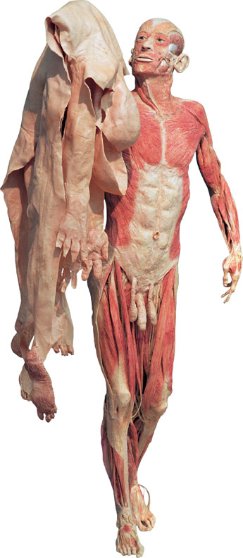
In a previous post we discussed how restrictions within the soft tissue surrounding the ankle joint may limit the ever so important motion of dorsiflexion.  However, soft tissue dysfunction or tissue adhesions may not be the only limitation when it comes to you and your ankle joint moving freely.  In fact, many times a gross reduction in motion may be caused by only a miniscule restriction in one of the surrounding foot and ankle bones, especially after injury.

This concept of “positional faults” was first made famous by Australian physiotherapist, Brian Mulligan in which he described an actual anterior translation of the fibula on the tibia after the typical inversion ankle sprain, rather than a disruption of the ATF ligament which so many athletic trainers and physical therapists focus their post-injury treatment protocol on.


Mulligan


“I believe that when the foot is forcibly plantar flexed and inverted beyond the natural range the lateral ligament usually suffers minor injury. The fibula gets wrenched forward on the tibia and positional faults occur.” (pg. 96 Manual Therapy by Brian Mulligan)


Mounting evidence is beginning to support this notion in some patients after the common ankle sprain as well as those with lateral ankle instability as indicated by the article summaries below.  As with all your patient cases, a proper and  methodical assessment is critical to determining if in fact a “positional fault” has occurred versus the previously discussed restriction within the soft tissue surrounding the ankle complex .  In the event that a patient has difficulty after a “simple” ankle sprain, consideration should be given to Mulligan’s concepts with manual therapy techniques employed to correct these faults.


Fibular Position in Individuals with Self-Reported Chronic Ankle Instability

by Hubbard, Hertel and Sherbondy


It has been reported that 55% of individuals who sprain their ankle do not seek treatment and up to 70% suffer from repetitive sprains. Chronic Ankle Instability (CAI) may also lead to increased risk of osteoarthritis and articular degeneration. “Altered arthrokinematics lead to abnormal physiologic motions, distorted state of ligamentous structure and altered joint function” (pg.4), with the combination of these possibly leading to an increased risk of re-injury.

Purpose: The purpose of this study was to assess the position of the distal fibula in individuals with chronic ankle instability CAI. 
Study Design: Thirty subjects with unilateral CAI and 30 subjects with no previous history of ankle injury participated in this study, completing a pair of subjective functional scales and fluoroscopic lateral images for both ankles with the distance from the anterior margin of the distal tibia to the anterior margin of the distal fibula measured.
Results: There were statistically significant differences in fibular position for the subjects with unilateral CAI compared to their non-injured limb as well as the control group, suggesting an anterior positional fault was present in those with unilateral CAI. 
Conclusion: the fibula was positioned significantly more anterior in relation to the tibia in subjects with unilateral CAI but further research is needed to determine if repetitive bouts of ankle instability caused the anterior fibula position or if the position was a predisposing factor to injury.
______________________________________________________________________________
Clinical Pearl: Suspicion of fibula position fault should be high in lateral ankle sprains with longer than usual symptoms despite traditional treatment, especially in those ankles with swelling that remains despite treatment and is supported by Hubbard and Hertel 2008 in which they found, “a strong positive correlation between fibular position and swelling.” In addition, “those ankles with more swelling had the most anteriorly positioned fibulae. The fibulae in sub-acutely sprained ankles appear to be positioned more anteriorly compared to the contralateral ankles. This positional fault may be maintained acutely by swelling.” (pg. 63)
______________________________________________________________________________


Immediate Effects of Anterior-to-Posterior Talocrural Joint Mobilization after Prolonged Ankle Immobilization: A Preliminary Study

by Landrum, Brent, Kelln, Parente, Ingersoll and Hertel


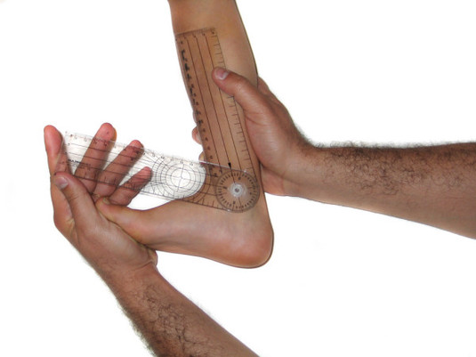
Ankle dorsiflexion ROM typically decreases after prolonged immobilization and thus is a central focus for many clinicians during the rehabilitation process.  Interventions such as static stretching and joint mobilizations are often employed as an effort to restore this very motion. “Passive mobilizations consist of gentle oscillating movements of the articular surfaces that create the movement of joints by a means other than the musculotendinous units that normally act on those particular segments. Joint mobilizations are purported to relieve pain and improve range of motion of injured joints.  Such  improvements can in turn lead to an increase in functional activities.” pg. 100. 


Purpose: “The purpose of this study was to determine if a single bout of Grade anterior-to-posterior talocrural joint mobilizations immediately affected measures of dorsiflexion ROM, posterior ankle joint stiffness, and posterior talar translation in ankles of patients who have been immobilized at least 14 days.”
Study Design: The study used 10 subjects 5 male and 5 female each who had been previously immobilized following lower extremity injuries with at least a 5 degree deficit in ankle dorsiflexion. Immobilization ranged from 2 weeks to 9 weeks. All subjects underwent three series of measurements of ankle dorsiflexion ROM, posterior talar mobility, and posterior joint stiffness. ROM was measured using a bubble inclinometer and talar mobility and joint stiffness were measured using an ankle arthrometer. The subjects were split into two groups one receiving the intervention and one control group. All subjects received both the joint mobilization and control (no intervention) in their crossover design.
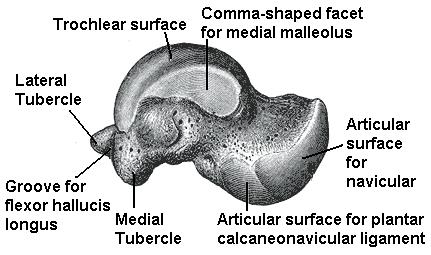
Results: Ankle dorsiflexion ROM increased significantly at each assessment period. A possible reason for this increase in dorsiflexion ROM could be due to correction of an anterior positional fault of the talus after joint mobilizations. It is possible that these faults were corrected via either the joint mobilization and/or the arthrometer testing.
Conclusion: “After a single application of Grade III anterior-to-posterior talocrural joint mobilization, dorsiflexion ROM and posterior ankle joint stiffness were significantly increased.  There was also a trend toward less posterior talar translation immediately after immobilization.”  This result may be related to correction of a positional fault at the talocrural joint.

______________________________________________________________________________
Interesting Note: “Mulligan’s positional fault theory may help explain our findings. Residual loss of the posterior glide may be representative of an anterior positional fault of the talus on the tibia and may result in an abnormal axis of talocrural rotation.  Through an acute mechanism of injury, such as ankle sprain, the talus may anteriorly subluxate and become stuck, thus resulting in restricted posterior glide and compromised ankle function.  It is possible that the patients in our study who were immobilized for a prolonged period of time also developed positional faults of the talus and that these positional faults were corrected via either the joint mobilizations and/or the arthrometer testing.” pg. 104
______________________________________________________________________________


Predicting Short-Term Response to Thrust and Nonthrust Manipulation and Exercise in Patients Post Inversion Ankle Sprain

By Witman et al.


“It has been estimated that the reinjury rate following a lateral ankle sprain may be as high as 80% suggesting the need to identify the most effective management strategies for this condition.”


Purpose: The purpose of this study was to develop a clinical prediction rule (CPR) to identify patients who had sustained an inversion ankle sprain who would likely benefit from manual therapy and exercise.
Study Design: Consecutive patients with a status of post inversion ankle sprain underwent a standardized examination to determine baseline data followed by a treatment program consisting of ankle/foot thrust and non-thrust manipulation, general mobility exercises (including ROM and stretching), advice to maintain usual activity within the limits of pain, and instruction in the use of ice and elevation. Thrust manipulations included a rearfoot distraction technique and a proximal tibiofibular joint posterior-to-anterior thrust manipulation. Nonthrust manipulation techniques used included anterior-to-posterior talocrural technique, lateral glide/eversion rearfoot technique and a distal tibiofibular technique. Of the 85 patients enrolled in the study, 64 or 75%, experienced a successful outcome as reported on the Global Rating of Change scale. Of those who experienced success, 35 or 55%, experienced a successful outcome at the time of the second visit and the remaining 29 experienced success at the 3rd visit. 
Conclusion: The study developed a CPR to identify patients with a status of post inversion ankle sprain who would most likely benefit rapidly and dramatically from manual therapy and general exercise. If 3 of the 4 variables (symptoms worse when standing, symptoms worse in the evening, navicular drop >5 mm, or distal tibiofibular joint hypomobility), were present, the accuracy of the rules was maximized and the post-test probability of success increased to 95%. 
______________________________________________________________________________
Interesting Note: Athletes/patients who have suffered ankle sprains in high school and did not seek medical help but instead left their ankle trauma to resolve on its own, often present years later with limited ankle range of motion with associated knee or other foot pain for which they usually present.  It is this author’s experience that athletes with previous ankle sprains, but otherwise “healthy” benefit greatly from the following mobilization techniques (described in this article) as part of their comprehensive treatment in an effort to regain normal ankle dorsiflexion regardless of their initial presentation. 


Appendix A – page 199

• Lateral Glides and Eversion Mobilization/Manipulation to the Subtalar Joint
• Talocrural joint Anterior-to-Posterior mobilization/manipulation with patient supine and ankle off end of treatment table
• Talocrural joint Anterior-to-Posterior mobilization/manipulation (mobilization with movement utilizing belt for assistance)


** This is true for both acute ankle inversion sprains as well as for athletes with residual restrictions from prior injury.  In a study by Green et al., in 2001 the addition of talocrural mobilizations to traditional RICE protocols necessitated fewer treatments to achieve pain-free dorsiflexion.
______________________________________________________________________________


Initial Changes in Posterior Talar Glide and Dorsiflexion of the Ankle After Mobilization With Movement in Individuals With Recurrent Ankle Sprain

By Vicenzino et al.


“Physiotherapists frequently use mobilization with movement (MWM) techniques as a physical treatment to improve range of motion, alleviate pain, and promote earlier return to function following lateral ankle sprain.”

Purpose: Test current contention that mobilization with movement (MWM) to the talocrural joint in both weight bearing and non–weight bearing positions improve posterior talar glide as a means to increasing ankle dorsiflexion
Study Design:  8 females and 8 males ages 18-27 recruited from Queensland’s student population
• Inclusion Criteria: “history of recurrent ankle sprain with at least 2 ankle sprains, more than 20 mm asymmetry on the weight-bearing lunge test for ankle dorsiflexion, no history of lateral ankle sprain on the contralateral side, and not receiving any other physiotherapy treatment during the study”
• Exclusion Criteria: “acute ankle sprain within past 6 months, previous injury or surgery in the back, hip or knee, or major fracture to the ankle or distal leg”

Results:  “weight-bearing and non-weight-bearing MWM treatment techniques both produced significant changes in posterior talar glide that were not evident in the control condition”

- Non-weight-bearing: “reduced posterior talar glide deficit by 50%”
- Weight-bearing: reduced posterior talar glide deficit by 55%

- Control: “reduced posterior talar glide deficit by 17%”
- Weight-bearing and nonweight-bearing MWM treatment improved weight-bearing dorsiflexion by 26%, compared to 9% by the control group.

Conclusion: “application of MWM treatment techniques improved posterior talar glide and talocrural dorsiflexion immediately after application in subjects with chronic recurrent lateral ankle sprain.”

____________________________________________________________________

Note:
“Approximately 44% of all sprained ankles go on to have further problems and although the factors that predispose to injury or reinjury are not conclusively evidence based, they are reported to involve proprioceptive deficits of the ankle, lack of ankle dorsiflexion, and reduced posterior glide of the talus in the ankle mortice.” (pg. 465)

“Denegar et al found a reduction in posterior glide of the talus in the ankle mortise in asymptomatic fully functional subjects in the 6 months following ankle sprain. It was postulated that because the talus lacks muscular attachments, it might subluxate anteriorly following disruption to the ligaments that attach to it. The talus then remains malpositioned anteriorly until it is passively returned to its ‘normal’ position. To an extent, the findings of reduced posterior talar glide and dorsiflexion range of motion appear congruent, as posterior talar glide is an accessory motion component of ankle dorsiflexion.” (pg. 465)
______________________________________________________________________________


Conclusion: Whether you are treating an acute ankle sprain, or addressing knee pain secondary to restricted ankle dorsiflexion a detailed evaluation which involves ALL contributing factors, both bony and soft-tissue, associated with a decrease in ankle dorsiflexion as outlined here and our previous post is the only way to ensure your patient will receive the best possible outcome and the fastest track back to normal function and high performance activities.  Positional faults are impossible to find if you never look for them - assessment of all ankle sprains or ankles with limited motion should include a detailed examination of all bones related to the foot and ankle including the distal tib-fib joint and talus for appropriate accessory motions. In a future post I’ll discuss why the all so common,  “ice and e-stim approach” to ankle sprain management only works for so long and why hip strength may actually be more important.


References

Collins N, Teys P, Vicenzino B. The initial effects of a Mulligan’s mobilization with movement technique on dorsiflexion and pain in subacute ankle sprains. Manual Therapy 2004; 9:77-82

Denegar, C., Hertel, J., Fonesca, J.  The Effect of Lateral Ankle Sprain on Dorsiflexion Range of Motion, Posterior Talar Glide, and Joint Laxity.  J Orthop Sports Phys Ther. 2002; 32(4):166-173.

Hubbard T, Hertel J, Sherbondy P. Fibular position in individuals with self-reported chronic ankle instability. J Orthop Sports Phys Ther. 2006;36(1):3-9.

Hubbard TJ, Hertel J. Anterior positional fault of the fibula after sub-acute lateral ankle sprains. Manual Therapy.  2008;13:63-67.

Landrum E, Kelln B, Parente W, Ingersoll C, Hertel J. Immediate effects of anterior-to-posterior talocrural joint mobilization after prolonged ankle immobilization: a preliminary study. J Manual Manip Ther. 2008;16(2):100-105.

Whitman J, Cleland J, McPoil T, et al. Predicting short-term response to thrust and nonthrust manipulation and exercise in patients post inversion ankle sprain. J Orthop Sports Phys Ther.  2009;39(3):188-200.

Vincenzino, B., Branjerdporn, M., Teys, P., Jordan, K.  Initial Changes in Posterior Talar Glide and Dorsiflexion of the Ankle after Mobilization With Movement in Individuals with Recurrent Ankle Sprain.  J Orthop Sports Phys Ther. 2006;36(6):464-471.

Green., T., Refshauge, K., Crosbie, J., Adams, R.  A randomized controlled trial of a passive accessory joint mobilization on actue ankle inversion sprains. Phys Ther. 2001;81(4):984-994.

Mulligan BR. Manual therapy: “NAGS,” “SNAGS,” “MWMS,” etc. 3rd ed. Wellington, New Zealand: Plane View Services LTD; 1995.


Topics: Basketball Related, Art Horne