Boston Sports Medicine and Performance Group, LLC Blog

Predicting Performance and Injury Resilience in Collegiate Basketball Athletes

Posted by Boston Sports Medicine and Performance Group on Wed, Aug 1, 2012 @ 06:08 AM


by Art Horne


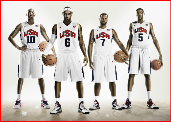
basketball performance resized 600


Just recently Dr. Stuart McGill, Jordan Andersen and myself published an article in the Journal of Strength and Conditioning Research examining the link between traditional pre-season strength, fitness, and sports medicine testing to overall on-court basketball performance and injury resilience throughout the course of two collegiate basketball seasons.  Although I would be the first to admit that there are some clear limitations to this study (number of participants for example), key performance predictors (points scored, ability to rebound, block shots, etc) were NOT associated with traditional strength or performance measures so often pursued in collegiate basketball strength programs.

Over the course of the next few weeks I will review this article in detail and provide insight into how actual on-court basketball performance may be improved upon beyond simply finding better parents or recruiting.  


Predicting Performance and Injury Resilience From Movement Quality and Fitness Scores in a Basketball Team Over 2 Years

McGill, Stuart M.1; Andersen, Jordan T.1; Horne, Arthur D.2

Journal of Strength and Conditioning Research

July 2012


Introduction

The ability to successfully predict injury resilience and competition performance from preseason testing is a very wishful goal; however, questions remain regarding this objective: Do tests of fitness have a predictive ability for injury and are there other factors that can be assessed that may predict injury? Are there specific indicators that predict performance? This study was motivated by these questions.

Attempts to understand injury mechanisms and performance sometimes consider links to fitness. Traditionally, fitness testing, at least in occupational settings, has included the assessment of strength (13), joint range of motion (ROM) (23), and physiological variables such as heart rate, blood pressure, and oxygen uptake (2), but the performance scores in the occupational context are difficult to quantify. In contrast, there have been some studies relating fitness to sporting performance that are more tangible. In studies of ice hockey players (6,24), success could be more tangibly quantified from on-ice measures such as total minutes played and scoring chances. Green at al. stated that “goals scored” was not the best measure of hockey skill. Studies of football players suggest that those who score higher on movement quality tests have few injuries (11,12); however, preseason football combine testing is dominated by tests of strength and running speed. Recognizing that movement asymmetry and compromises to neuromuscular control have been linked to both future injury (11,12) and with having a history of back injury (17), movement assessments have been developed (3,4) and have been suggested to predict injury rates. Further, several fitness and movement tests have been implicitly assumed to predict “playing” performance by their inclusion into standard preseason tests. These include tests of endurance, strength, joint ROM, agility, and speed. The question remains as to the validity of these factors when attempting to predict injury resilience and performance.

Although links between moving well and injury resilience and performance seem intuitive, this notion remains controversial. Interestingly, some evidence suggests that fitness training alone may not ensure peak performance or injury resilience (8,20). In addition, movement quality has been suggested to predict future injury (12). A possible mechanism may be that injury changes the way a person moves as an accommodation to pain (consider, e.g., the changes in mechanics throughout the anatomical linkage when limping from foot pain). Having a history of injury, in particular back injury, appears to change movement patterns (17). Movement patterns determine important injury criteria, such as joint and tissue load, together with influencing the length of time and repetitions an individual is able to perform a task with uncompromised form. Compromised form exposes the tissues to inordinate load elevating the risk of injury. Several examples of this link are available, for example, not maintaining a neutral curve in the lumbar spine while bending and lifting decreases the tolerable load at injury (in this case tissue failure [18]); having restricted hip motion is linked to having more spine motion when bending (17). Movement competency has also been linked with anterior cruciate ligament (ACL) injury rates, for example, having larger knee abduction moments and angles when landing from a jump predicted higher ACL injury rates (9). Given the variety of considerations for interpreting the links between movement, fitness, performance, and potential injury, the goal of this study was to first evaluate some traditional fitness test scores in a controlled athletic group that has a variety of challenging movement demands and also perform an assessment of the quality of movement. It was hoped that following a test group for a period of time would reveal links between specific fitness scores and movement quality with variables to predict injury resilience and performance. If such links exist, they could form a rationale for specific tests to be included in preseason testing.

The purpose of this study was to see if specific tests of fitness, and movement quality, could predict injury resilience and performance in a team of basketball players over 2 years (playing seasons).

It was hypothesized that in a university basketball population, (a) Preseason movement quality and fitness scores would predict in-season performance scores. (b). Preseason movement quality and fitness scores would predict in-season injury resilience.


See Dr. Stuart McGill and other world authority in Sports Medicine, Science and Performance at the 2013 BSMPG Summer Seminar - May 17 & 18 in Boston MA

Topics: Art Horne, Brian McCormick, basketball performance, basketball conference, basketball training programs, athletic training conference, Craig Liebenson, Shawn Windle, Basketball Training, Stuart McGill, Keith D'Amelio

Leopards or Turtles? by Jose Fernandez

Posted by Boston Sports Medicine and Performance Group on Fri, May 11, 2012 @ 07:05 AM

BSMPG


Basketball is a multifactorial sport where recovery, nutrition, training, technical & tactical aspects, mental preparation and innate conditions are involved. As S&C coaches, our ultimate goal is to enhance the team performance by optimising each player´s physical condition and helping them stay away from injuries.

Profiling athletes is an important part of the training process that helps me to decide what is the most appropriate strategy for each of the players I coach.

The image below represents the average results of 3 pre-season assessments to determine the % of Type I muscle fiber (Slow Twitch). It is an example of two different football players, both of them playing for the same team but with a different muscular profile.

The player on the left seems to have lower predominance of slow twitch as every muscle group except Semitendinosus (very postural muscle) is within 30-45% of slow muscle fibres.

The player on the right seems to have higher predominance of slow twitch, especially on key muscle groups like Biceps Fem (59,8%) and Gluteus Max (62%).

Click HERE to continue reading...

See Jose and other internationally known speakers at the 2012 BSMPG Summer Seminar May 19-20th.

Hurry - Seats are limited.


Click me


Topics: Art Horne, basketball performance, BSMPG, Craig Liebenson, boston hockey conference, Cal Dietz, Bill Knowles, Alan Grodin, Barefoot in Boston, Dan Boothby

Up the Chain It Goes... (Part II)

Posted by Boston Sports Medicine and Performance Group on Fri, May 4, 2012 @ 07:05 AM

By Art Horne


turf toe derrick rose toe injury


In a follow up from a previous post (Up The Chain It Goes), additional evidence supporting the relationship within the kinetic chain has emerged from south of the equator.  In a study out of South Africa examining the link between available dorsiflexion and mechanical low back pain researchers found a statistically significant decrease in ankle dorsiflexion ROM and associated reporting of low back pain (Brantingham, 2006).   With the vast majority of adults suffering from low back pain at some time in their life, (some reports are up to 85%) and 80% of people reporting foot problems during their lifespan, it’s not a surprise to see that these two conditions may very well be related.

Let’s take a closer look:

Methods: “ The study was a blinded, 2-arm, non-randomized clinical study involving 100 subjects with chronic or recurrent mechanical low back pain (intervention group) and 104 subjects without chronic mechanical low back pain (control group) between the ages of 18 and 45.  A blind assessor performed weight-bearing goniometry of the ankle and big toe and the navicular drop test on all subjects in both groups.”

Results: “An independent t-test (inter-group) revealed a statistically significant decrease in ankle dorsiflexion range of motion in individuals with chronic mechanical low back pain.”

Conclusions: “This study’s data found that a statistically significant decrease in ankle dorsiflexion ROM, but not flatter feet, was associated with subject report of chronic mechanical low back pain disorders.”

Discussion: “The findings of this blinded study support previous reports suggesting that decreased ankle dorsiflexion may be a factor in chronic mechanical low-back pain.  There was no clear association found between decreased hallux ROM and mechanical low back pain in this study.  If these findings are confirmed through additional studies, exercise and manipulation therapy to increase ankle range of motion could become an important consideration in the treatment of some patients with mechanical low back pain disorders.”

Hmmm, if only we had some additional studies….

Perhaps this will help.

During a routine exit physical, 60 division one athletes were assessed for available weight bearing dorsiflexion bilaterally as described by Bennell et al in 1998 (inclinometer was replace by Clinometer app for ITouch) to examine limitations in this movement.   Ten athletes with limited weight bearing dorsiflexion (less than 4 inches from knee to wall) volunteered for follow up evaluation and manual treatment. Out of the initial 120 measured ankles, 47 ankles (21 right, 26 left) demonstrated limited weight bearing dorsiflexion range of motion.

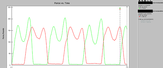
Athletes were then asked to walk normally in their athletic shoes while wearing an in-shoe pressure sensor (Tekscan) and through an optical measurement system (Optojump).  Each athlete then underwent a general manual therapy intervention aimed to improve ankle dorsiflexion, followed again by the same gait analysis and pressure mapping data capture.


Gait Evaluation


Gait Cliff Notes: optimal gait should have two mountains with a trough between them. The first mountain represents heel strike to midstance, the trough representing the mid-stance phase, and the second mountain being propulsion from full foot contact to toe-off.

Easy right? Good. 

Note: The second mountain should almost always be higher than the first.


Case Study 1:

Tekscan report


Pre-treatment (RED):

Notice how the first mountain is slightly higher than the second – this is BAD!

Remember from our cliff notes: the second mountain should be higher.

Post-treatment (GREEN):

Notice change in toe off from pre- to post-treatment which specifically targeted patient's limited dorsiflexion?  The second mountain is now higher than the first. That’s a GOOD thing!

Awesome right?

Better yet – athlete was measured 3 days post treatment and improvement in Dorsiflexion range of motion stuck!  Try doing that with a slant board stretch.


See Art Horne and Dr. Pete Viteritti discuss these and other changes at the foot and ankle, and how to assess and address soft tissue and bony restrictions in their presentation at the 2012 BSMPG Summer Seminar May 19-20th in Boston.


Click me


References

Bennell KL, Talbot RC, Wajswelner H, Techovanich W, Kelly DH and Hall AJ. Intra-rater and inter-rater reliability of a weight-bearing lunge measure of ankle dorsiflexion. Australian Journal of Physiotherapy. 44;175-180.

Brantingham JW, Gilbert JL, Shaik J, Globe G. Sagittal plane blockage of the foot, ankle and hallux and foot alignment-prevalence and association with low back pain. J of Chiropractic Medicine. 2006; 4(5); 123-127.


Topics: Art Horne, basketball performance, basketball training programs, boston hockey conference, barefoot strength training, Bruce Williams, Cal Dietz, Bill Knowles, Barefoot in Boston, Chris Powers, Dorsiflexion

Up the Chain it Goes...

Posted by Boston Sports Medicine and Performance Group on Sun, Apr 29, 2012 @ 15:04 PM

By Art Horne


derrick rose torn acl


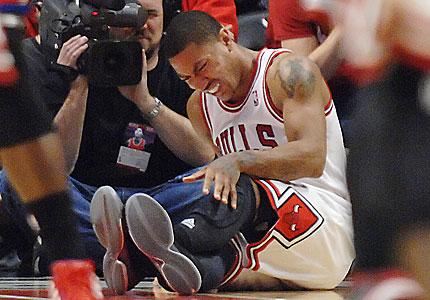
With recent season ending ACL injuries to New York Knicks Iman Shumpert, and Chicago Bull’s point guard Derrick Rose coming on the same day, (not to mention Eric Maynor from the Thunder and Spanish Star Ricky Rubio earlier this season) discussion has arisen as to how these terrible injuries could have been avoided.  Although the possible contributing factors are endless, ranging from previous injury to simply fatigue, one area worth shedding more light on, especially in the case of young Rose, is the implication of the kinetic chain as a whole.

Let’s start at the ground and work our way up.

I think we’d all agree that the big toe is a big deal.   But how closely are we looking at this “pivotal” body-ground juncture?

In a study by Munuera et al, researchers found that “Hallux interphalangeal joint dorsiflexion was greater in feet with hallux limitus than in normal feet.  There was a strong inverse correlation between first metatarsophalangeal joint dorsiflexion and hallux interphalangeal joint dorsiflexion.” (Munuera et al, 2012). 

TRANSLATION: People with abnormally stiff or limited motion at the great toe had excessive motion at the joint just distal.

If you don’t have mobility where you need it, you’ll surely get it somewhere else.

Let’s move up the chain shall we?

In a study by Van Gheluwe and his group,  researchers looked at how a stiff or limited great toe joint changes the way we walk.  In their study, “two populations of 19 subjects each, one with hallux limitus and the other free of functional abnormalities, were asked to walk at their preferred speed while plantar foot pressures were recorded along with three-dimensional foot kinematics.  The presence of hallux limitus, structural or functional, caused peak plantar pressure under the hallux to build up significantly more and at a faster rate than under the first metatarsal head.  Additional discriminators for hallux limitus were peak dorsiflexion of the first metatarsophalangeal joint, time to this peak value, peak pressure ratios of the first metatarsal head and the more lateral metatarsal heads, and time to maximal pressure under the fourth and fifth metatarsal heads.  Finally, in approximately 20% of the subjects, with and without hallux limitus, midtarsal pronation occurred after heel lift, validating the claim that retrograde midtarsal pronation does occur.”

TRANSLATION: if you have a limited motion in your great toe, pressure changes will occur – increase pressure changes will cause pain over time (think blister on your foot).

And pain changes the way we move – period.

Let’s take a look at the ankle.

In an article  by Denegar et al, the authors outline the importance of regaining normal talocrural joint arthrokinematics following an ankle injury.   The authors note,

 “All of the athletes we studied had completed a rehabilitation program as directed by their physician under the supervision of a certified athletic trainer, and had returned to sports participation.  Furthermore, all had performed some form of heel-cord stretching. None, however, had received joint mobilization of the talocrural complex.  Despite the return to sports and evidence of restoration in dorsiflexion range of motion, there was restriction of posterior talar mobility in most of the injured ankles.  Posterior talar mobilization shortens the time required to restore dorsiflexion range and a normal gait.  Without proper talar mobilization, dorsiflexion range of motion may be restored through excessive stretching of the plantar flexors, excessive motion at surrounding joints, or forced to occur through an abnormal axis of rotation at the talocrural joint.” (pg. 172)

TRANSLATION: I repeat, Without proper talar mobilization, dorsiflexion range of motion may be restored through excessive stretching of the plantar flexors, excessive motion at surrounding joints, or forced to occur through an abnormal axis of rotation at the talocrural joint.” (pg. 172)

If you don’t have normal ankle motion, and specifically at the talus, your ankle motion (although appearing normal) is probably coming from other joints and/or in a combination with foot pronation.


Foot Pronation = Tibial Internal Rotation

Tibial Internal Rotation = Femoral Internal Rotation

Tibia and Femur Internal Rotation  =  Knee Valgus (or knee collapse)

Knee Valgus = BAD


But just because you have some extra motion doesn’t mean you’re doomed right?

No.

But, excessive motion without the ability to control that motion certainly does.  So where does knee control come from? The Hip!

But hip strength, control, and neuromuscular timing is seldom appreciated, and in the case of the basketball athlete it is certainly poorly measured, especially after ankle injury.

In a study by Bullock-Saxton, researchers investigated muscle activation during hip extension after ankle sprain and showed a changes in timing of muscle activation in the ankle sprain grouped compared to the non-injured group.

 “the results highlight the importance of the clinician’s paying attention to function of muscles around the joints separated from the site of injury.  Significant delay of entry of the gluteus maximus muscle into the hip extension pattern is of special concern, as it has been proposed by Janda that the early activation of this muscle provides appropriate stability to the pelvis in such functional activities as gait.” (pg. 333)


In another study examining ipsilateral hip strength/weakness after the classic ankle sprain, researchers demonstrated that subjects with unilateral chronic ankle sprains had weaker hip abduction strength and less plantar flexion range of motion on the involved sides (Friel et al., 2006)

“Our findings of weaker hip abductors in the involved limb of people with chronic ankle sprains supports this view of a potential chronic loss of stability throughout the kinetic chain or compensations by the involved limb, thus contributing to repeat injury at the ankle.” (pg. 76)


“If the firing, recruitment, and strength of the hip abductor muscles in people with ankle sprains have been altered because of the distal injury, the frontal-plane stability normally supplied by this muscle is lacking, and the risk for repeat injury increases.  Weak hip abductors are unable to counteract the lateral sway, and an injury to the ankle may ensue.”

TRANSLATION: Ankle sprains cause neuromuscular changes up the chain and specifically in the hip.  If this weakness is not addressed after an ankle injury,” frontal-plane stability normally supplied by this muscle is lacking.” 


Lack of frontal-plane stability + Knee Valgus = Injury


Of course suggesting that the above points are exactly the reason for which Rose suffered his injury is certainly a stretch and not the intention of this post, nor is it to question the treatment that he or any other NBA player received prior to their devastating injury (for the record, the Chicago Bulls Sports Medicine and Strength Staff are regarded as one of the very best in the league).  What I am suggesting however is that examining athletes and patients with the use of advanced technology to determine a state of readiness to participate, and/or examine more closely changes in gait and neuromuscular firing is certainly worth pursuing, especially in light of the ever-rising salaries within professional sports.  A quick look is certainly worth the small investment.

One thing is for sure, ACL injury is not limited to teenage females or only seen on the soccer pitch.


Previous Posts:

The NBA Should Have Learned From The NFL - Injuries On The Rise

Did The NBA Lock-out Ultimately End Chauncey Billups' Career?


See lectures directly related to gait, injury prevention, and performance at the 2012 BSMPG Summer Seminar:

1. Dr. Bruce Williams: Hit the ground running: Appreciating the importance of foot strike in NBA injuries

2. Dr. Bruce Williams: Breakout Session: Restoring Gait with evidence based medicine

3. Art Horne and Dr. Pete Viteritti: Improving Health & Performance - Restoring ankle dorsiflexion utilizing a manual therapy approach

4. Dr. Tim Morgan: Biomechanics and Theories of Human Gait: Therpeutic and Training Considerations

5. Jose Fernandez: Advanced Player Monitoring for Injury Reduction


See the most advanced player monitoring equipment currently available at the 2012 BSMPG Summer Seminar:


 zeo affectiva  ithlete

BioSensics  Zflo insideTracker


Dartfish  freelap timing   Tekscanoptosource

Click me


References:

Munuera PV, Trujillo P, Guiza L, Guiza I. Hallux Interphalangeal Joint Range of Motion in Feet with and Without Limited First Metatarsophalangeal Joint Dorsiflexion. J Am Podiatr Med Assoc. 102(1): 47-53, 2012.

Denegar, C., Hertel, J., Fonesca, J.  The Effect of Lateral Ankle Sprain on Dorsiflexion Range of Motion, Posterior Talar Glide, and Joint Laxity.  J Orthop Sports Phys Ther. 2002; 32(4):166-173.


Van Gheluwe B, Dananberg HJ, Hagman F, Vanstaen K. Effects of Hallux Limitus on Plantar Foot Pressure and Foot Kinematics During Walking. J Am Podiatr Med Assoc. 96(5): 428-436, 2006.

Bullock-Saxton, J. E., Janda, V., & Bullock, M. I. (1994) The Influence of Ankle Sprain Injury on Muscle
Activation during Hip Extension. Int. J. Sports Med. Vol. 15 No. 6, 330-334.

Friel, K., McLean, N., Myers, C., & Caceres, M. (2006). Ipsilateral Hip Abductor Weakness After Inversion
Ankle Sprain. Journal of Athletic Training. Vol. 41 No.1, 74-78

Smith RW, Reischl SF. Treatment of ankle sprains in young athletes. Am J Sports Med. 1986;14:465-471.

Topics: Art Horne, basketball performance, basketball training programs, BSMPG, athletic training conference, Charlie Weingroff, boston hockey conference, barefoot strength training, Andrea Hudy, Bruce Williams, Cal Dietz, Alan Grodin, Barefoot in Boston, Dr. DiMuro, Dan Boothby, Chris Powers, achilles pain, Dorsiflexion, ankle problems

Kansas' Secret Weapon - Andrea Hudy Comes to Boston

Posted by Boston Sports Medicine and Performance Group on Wed, Apr 4, 2012 @ 06:04 AM

Andrea Hudy

University of Kansas Strength & Conditioning Coach

Andrea Hudy


When the 7-foot center Jeff Withey showed up on the Kansas campus in 2009, he was a gawky San Diego kid who weighed a shrimp taco or two above 200 pounds. So how did he develop into the bruiser who has helped put the Jayhawks into the NCAA tournament's Final Four?

Withey credits two people. The first is Kansas assistant coach Danny Manning, a Jayhawk legend who won the 1988 national title, was selected No. 1 in the NBA draft and recently was named Tulsa's new coach. The other is a blonde-haired former college volleyball player named Andrea Hudy.

Withey describes her as "one of our secret weapons."

Click HERE to continue reading.


See Andrea and other top basketball coaches from across the country speak at the BSMPG Summer Seminar - May 19/20 in Boston.


Click me

Topics: basketball performance, basketball conference, athletic training conference, Craig Liebenson, Andrea Hudy, Cal Dietz, Bill Knowles

Advanced Athlete Monitoring For Injury Reduction at the 2012 BSMPG Summer Seminar

Posted by Boston Sports Medicine and Performance Group on Wed, Mar 7, 2012 @ 07:03 AM

Join Jose Fernandez and other top Sports Medicine and Sports Performance professionals from across the world as they desend on Boston this May 19th-20th for the 2012 BSMPG Summer Seminar.  Choose from a number of specific learning tracks or mix and match to suit your learning needs.  Keynote speakers throughout the weekend include Dr. Craig Liebenson, Chris Powers, Alan Grodin, Irving Schexnayder, and Bill Knowles. 

Register today for this once in a lifetime event! Seats are limited!


BSMPG Summer Seminar


        BSMPG Summer Seminar


Boston Sport Medicine and Performance Conference

Advanced Athlete Monitoring for Injury Reduction

Jose Fernandez


Abstract:

Professional athletes are experts at what they do, regardless how many S&C sessions they perform a week, they either have the quality to average 20 points per night or they don´t. From a physical perspective, coaches need to make sure their athletes are healthy and available to play every night. A healthy professional athlete should be capable to display a good performance just by being healthy. Everything else that can be achieved with training is a plus.

In a league where teams have to play 3-4 games a week and take more than 90 flights per season, time is limited for coaches to carry out physical training sessions with their players. A training program must be precise, specific and adjusted to the individual needs of each athlete. Coaches should focus on maintaining and reducing the loss of training adaptations throughout the season while enhancing the recovery and regeneration strategies.

At this year´s BSMPG Conference, I will be presenting ideas on how to objectively profile athletes attending to their neuromuscular characteristics and type of muscle fiber predominance. Continuing with this neuromuscular approach to athlete monitoring, innovative ways to quantify effects and duration of the training and treatments will be discussed. Being able to control the rate at which each muscle gains and looses activation after a training session or how exactly certain therapy treatment affects the functionality of any muscle group is crucial if we want to schedule training actions at the right moment, with the aim to maximize the physical performance and minimize risk of injury during the competition.

From an injury prevention perspective, new concepts to evaluate athlete´s readiness to train and assessment of change in muscle response induced by training will also be suggested, with a clear focus on practicality and applicability.

Aiming for maximal performance is a complex task. The purpose of my presentation is to offer some insight into the analysis of an athlete´s neuromuscular condition and how this can help coaches optimize training in an objective, reliable and time saving manner.

I look forward to seeing you at the BSMPG Conference in May!


Topics: Art Horne, basketball performance, BSMPG, athletic training conference, boston hockey conference, Logan Schwartz, Andrea Hudy, Bruce Williams, Mark Toomey, Cal Dietz, Alan Grodin, Joel Jamieson, Dan Boothby, Jose Fernandez

Essential Reading Spotlight - Greenman's Principles of Manual Medicine

Posted by Boston Sports Medicine and Performance Group on Wed, Oct 12, 2011 @ 06:10 AM

I have to admit I missed the boat on the importance of manual medicine for a long time.  After speaking to the best clinicians and therapists in the area there was one book that each and everyone of them, regardless of their current treatment approach, recommended I read - Greenman's Principles of Manual Medicine (Point (Lippincott Williams & Wilkins))   

After recently completing this book I now know why.  This book is a must for anyone treating musculoskeletal dysfunction on a daily basis.  Below is a short excerpt from this book.


The Manipulable Lesion

"The acceptable term for this entity is somatic dysfunction. It is defined as impaired or altered function or related components of the somatic (body framework) system; skeletal, arthrodial, and myofascial structures; and related vascular, lymphatic, and neutral elements. Notice that the emphasis is on altered function of the musculoskeletal system and not on a disease state or pain syndrome." pg 11


DIAGNOSTIC TRIAD FOR SOMATIC DYSFUNCTION 


"The mnemonic ART can express the diagnostic criteria for identification for somatic dysfunction.

“A” stands for asymmetry of related of the musculoskeletal system, either structural or functional.
“R” stands for range of motion of a joint, several joints, or region of the musculoskeletal system. The range of motion could be abnormal by being either increased (hypermobility) or restricted (hypomobility). The usual finding in somatic dysfunction is restricted mobility, identified by observation and palpation using both active and passive patient cooperation.
“T” stands for tissue texture abnormality of the soft tissues of the musculoskeletal system (skin, fascia, muscle, ligament, etc.). Tissue texture abnormalities are identified by observation and a number of different palpatory tests.

Some authors add one of two other letters to this mnemonic. “P” or a second “T”. “P” stands for pain associated with other findings, and “T” stands for tenderness on palpation of the area. Tenderness is particularly diagnostic if localized to a ligament. A normal ligament is not tender. A tender ligament is always abnormal. However, both pain and tenderness are subjective findings instead of the objective findings of symmetry, altered range of motion, and tissue texture abnormality. By the use of these criteria, one attempts to identify the presence of somatic dysfunctions, their location, whether they are acute or chronic, and particularly whether they are significant for the state of the patients wellness of illness at that moment in time. In addition to the diagnostic value, changes in these criteria can be of prognostic value in monitoring the response of the patient, not only to manipulative treatment directed toward the somatic dysfunction, but also to other therapeutic interventions."  Pg 11-12


Topics: Art Horne, basketball performance, basketball resources, BSMPG, athletic training conference, boston hockey summit, boston hockey conference, barefoot strength training, barefoot training

Integrated Care - Part V: When NOT Touching Your Toes is NOT a Hamstring Problem

Posted by Boston Sports Medicine and Performance Group on Sun, Sep 18, 2011 @ 06:09 AM

by Art Horne


I remember being taught in school that people have low back pain because their hamstrings are tight.  Therego, stretch the hamstrings and resolve the low back pain.  It was a simple solution to a very complex and often misunderstood problem and yet as a student it was a clean and direct resolution to an often nagging dilemma.

Further testing and evaluation always “proved” the hamstrings as the culprit since so many of the low back pain patients were never able to touch their toes, and of course toe touching was a direct result of hamstring length.  Again, this bore out to make sense since after spending an exhausting amount of time stretching these patient’s hamstrings, and then retesting, some measurable change were noted (sometimes) with the patient inching closer to their toes.  The only problem in this clean and concise “hamstring-LBP” relationship was that these patients, no matter how long I stretched them or which stretching technique I employed ALWAYS ended up coming back the very next day for the same stretching routine and the same unresolved back pain.


BSMPG

Insanity: Doing the same thing over and over again and expecting different results.


Of course, many of these patients really never had “tight” hamstrings after all, but instead had hamstrings that were preventing them from a place they no had no business being in the first place. 

What do I mean?

Well, many of these patients, or at least the majority of them,  were flexion intolerant which means both flexing their spine and/or moving into flexion caused a reflexive “tightening” of the hamstrings to essentially keep them from FLEXING FORWARD and moving into a region which would exacerbate their current condition, or “a place they have no business being.”  In addition, many of these “tight hamstring” patients were never ever feeling a hamstring stretch, but instead were experiencing a neural stretch, which unfortunately after stretching only continued their “tight-stretch-pain-tight” cycle. 

Soo… Should I Stretch or Not?

1. First distinguish between hamstring tightness and neural tension.  Neural tension is always described differently than muscular tightness – that is if you’re listening.  I had a professor who told me that your patient will always tell exactly what is wrong with them if you listen long enough.  And if you listen just a little bit longer, they’ll also tell you exactly how to make them better.  This case is no different – of course, if you’re too busy catering water you’ll never have enough time to listen long enough.

2. Neural tension is often described as “pinching” or with other words that clearly denote nerve origin such as “zapping” or “burning.”  Muscle tension doesn’t zap or burn.

3. Confirm neural involvement with a slump test – then STOP stretching and begin a nerve flossing regiment if indicated.

4. Does the patient need more mobility (hamstring stretching) or do they simply lack the appropriate stability (or neuromuscular control) which ultimately is limiting them from moving into a place where they simply didn’t have the requisite control? I’m willing to bet that more often than not that stability/neuromuscular control is the limiting factor and not hamstring length – especially in low back pain patients.

So what does this have to do with integrated care?

1. On evaluation during pre-participation screenings a simply toe touch as a gross indicator of both ability and willingness to forward flex will “catch” those athletes who either have had previous back pain, current back pain or a complete lack of understanding relating to lumbar spine/pelvis position and an ability to disassociate them during normal daily living.

2. Those with pain or previous pain should be referred to sports medicine for further evaluation – yes, even those that don’t currently have pain.

3. Those that have trouble separating hip and lumbar motion should be placed into a “teaching” group while in the weight room until they can successfully “stiffen” their spine while gaining mobility and motion throughout the hips; both which are essential if any type of squat or deadlift pattern are prescribed in their program.  But really more importantly, these two patterns occur each and every day of their lives and must be grooved before major lumbar spine pathology presents itself.

Click HERE for a quick note on establishing a hip hinge.


Next week: A quick in-service to get both Sports Medicine and Strength Training hinging at the hips and teaching all athletes a great squat pattern. 

Topics: Art Horne, basketball performance, basketball resources, basketball conference, basketball training programs, athletic training conference, barefoot strength training, Barefoot in Boston, barefoot training

Integrated Care - Part III: Breaking Down Extension Based Problems

Posted by Boston Sports Medicine and Performance Group on Mon, Sep 5, 2011 @ 07:09 AM

by Art Horne


Although identifying a poorly executed squat is easy for many sports medicine and strength professionals (especially given the “rules” and guidelines set forth by the FMS standards), identifying the actual underlying problem or major contributor to a deficit squat is never as easy.  However, with a systems based approach during your annual pre-participation screenings your team of health care and performance professionals can tease out these often overlooked deficiencies as part of your movement screen from the very beginning instead of reacting after future injury or poor performance.

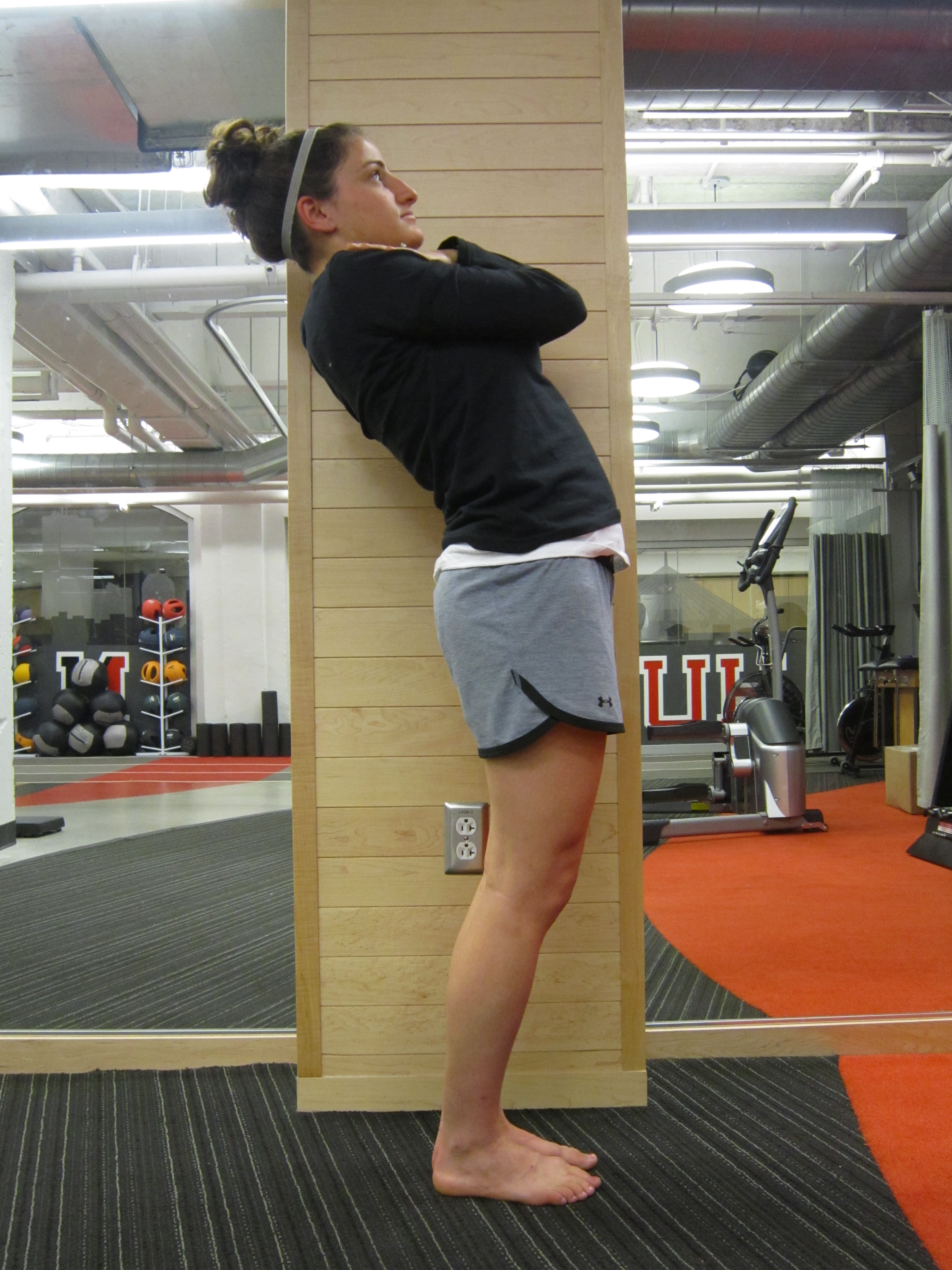
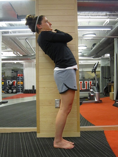
As I mentioned in a prior post the ability to express a proper squat pattern is fundamental to human movement.  Included in this category of fundamental “expressions” is the overhead reach, or what is commonly known through the Selective Functional Movement Screen TM (SFMA) as the Multi-segmental Extension Pattern (MSE) pictured below (figure1).  This easy test that takes literally 10 seconds can produce some startling results when athletes and those observing them, witness an otherwise “healthy”  and  highly skilled athlete in the prime of their life unable to reach behind them while shifting their weight and hips forward. 


functional movement screen

figure 1.


Let’s break it down.


According to the SFMATM rules, or simple human movement fundamentals, normal range of motion during this test includes:

1. With heels together (this is important and often overlooked as it provides a test-retest standardization) an athlete or patient should be able to have their ASIS pass over their great toes while;

2. Reaching overhead with hands in line with their shoulders and have the spine of their scapula clear or pass behind the heels of their feet and;

3. Their hands clear or pass behind the spine of the scapula (Figure 2).


functional movement

figure 2.


These movement minimums allow clinicians and performance coaches a starting point to begin observing their athletes for general restrictions within this pattern.  If an athlete is unable to complete this movement (failure to pass the ASIS over the great toe) then the athlete is asked to cross their arms over their chest and repeat the movement.  If the athlete is unable to still exhibit this fundamental expression of extension then the athlete in my opinion should be referred to a staff athletic trainer for further evaluation with the underlying problem is identified and addressed.  Many times the athlete will not understand why they are being referred since they have never had a “problem” before, but after a quick evaluation and treatment you’ll often find their eyes beam wide open with the additional range and freedom of motion that you’ve given back to them.

To confirm your findings on the examination table (although authentic human movement rarely ever happens on an exam table. Side note: How come you can measure with a goniometer an athlete’s ankle, knee and hip range of motion on an examination table and determine that they have the requisite mobility to perform a normal and unrestricted squat pattern but when you stand them up, the pattern looks like a train wreck? Movement means so much more than just your standard orthopedic examinations), have the athlete lay prone and ask them to tighten up one butt check and extend their leg off the table.  Repeat with the opposite leg and compare.  Rarely will an athlete be able to exhibit the appropriate amount of hip extension during this prone table test and not be able to pass their ASIS over their toes during the standing evaluation with arms crossed over head.

So what’s next? How do I fix it?

Not so fast.  I think it’s worth mentioning here a few items that support adding this simple test into both your yearly screenings along with your general orthopedic examination (regardless of injury presentation).  First off, I have been utilizing the FMSTM screen for a very long time as a strength coach, and over the last year have been utilizing the SFMATM methodology during injury presentation in the clinic and the overwhelming end result to many movement dysfunction and injury/pain cases have always boiled down to two movement impairments – Shoulder Mobility (Which in the end really is T-Spine Mobility) and Hip Extension.  The FMSTM  includes a test called the Active Straight Leg Raise, and this test unfortunately has been deemed a “hamstring” flexibility test or a hip flexion test by most casual observers but this couldn’t be further from the truth or the original intent of the test (future post coming: Are your hamstrings tight or are they just not letting you go somewhere you have no business being?).

For those that are not familiar with the test, an athlete lays on their back with a 2x6 board under their knees and while keeping the bottom leg in contact with the board slowly raises an extended leg upwards exhibiting the DIFFERENCE and available motion between the two legs and NOT the amount of hamstring or hip flexion range that you have.  It is this DIFFERENCE that should be noted, which ultimately leads to an examination and treatment focus of the down-leg in most instances as I mentioned earlier as the limiting factor (hip extension).

This should really come as no surprise since we have clearly become a hip flexion dominant society (sitting at computers, video games, etc not to mention our affinity for sitting on bikes at the commercial gym and watching the TV screen instead of sprinting on an incline treadmill which of course requires a bit of hard work and the aforementioned hip extension) and have basically lost the ability to “express” hip extension.  Although strength coaches and sports medicine professionals alike advocate “stretching” this problematic area after injury I think it’s worth teasing out your future patients sooner than later with a simple test while they are healthy athletes and avoid their inevitable future visit to your sports medicine clinic as patients.


Next week: Addressing and Correcting this Hip Extension Problem from both sides of the wall.


Topics: basketball performance, basketball training programs, BSMPG, FMS, SFMA, functional movement screen, Barefoot in Boston

Integrated Care - Assessment and Intervention

Posted by Boston Sports Medicine and Performance Group on Mon, Aug 29, 2011 @ 07:08 AM

by Art Horne


At the college level many times both assessment and intervention decisions are made based on time availability and simple manpower, and not on what the student-athlete requires for optimal health and performance.  Juggling study hall, practice, classes along with rehabilitation and performance training leaves little time for “additional” work for either the student-athlete or the staff professional in charge to provide additional auxiliary services in the form of corrective work, soft tissue manipulation or additional strength training.  With that said, this extra “work” is often neglected or pushed aside until either the student-athlete is no longer able to participate in practices or games due to an injury or becomes crippled due to some form of debilitating pain.  In either case, unfortunately the student-athlete has now become a student-athlete-patient within your facility and the little time you had to address her problem prior (which of course is why it wasn’t taken care of in the first place, or even looked at – ignorance is bliss after all) has now become a major investment and drain on your time and services. 

In order to avoid the initial trap that so many sports medicine and performance departments fall into each fall it is paramount that both departments (Sports Medicine and Strength Training) first reach an agreement to implement a comprehensive screening program TOGETHER to tease out dysfunction, evaluate for painful movement patterns and address these minor “tweaks” before they become major pains.


athletic training


Where to start:


It’s hard to rank which movement pattern is more important over another as each of the “Big Three” (squat, lunge and step) are all integrated and hold value within the context of all sporting events and training.  However, the only pattern among the three that is universally tested among college athletes and Strength Coaches is the squat, and thus, at least with regards to an integrated approach takes precedent over the others if having to choose only one.  Administering the test takes under a minute and produces so much more than just a number via the traditional FMS scoring system.


1. There’s something powerful about having members of both your sports medicine and strength staffs stand beside each other while evaluating a student-athletes overhead squat pattern during a fall pre-participation examination.  Because the strength coaches typically tests each athlete’s squat either later the same day or the next, this “pre-screening” allows strength coaches to see the movement pattern in an authentic form, not to mention in a rare one-on-one format which is never the case in a collegiate weight room due to traditional low coach to student ratios. 

2. When an athlete scores a “1” which means they cannot achieve a proper squat, it’s always nice to see the strength coach cross the name off the list of kids to max test later that day.  If you cannot squat to at least a “2” in the FMS overhead squat test then you simply haven’t qualified to load the pattern and go balls to the wall during testing – PERIOD.  This will sometimes be an issue among sports medicine professionals and strength coaches if the athletic trainer simply tells the strength coach that the athlete shouldn’t squat; but this is never a problem when the strength coach sees for themselves the awful pattern that the student-athlete exhibits.  The strength staff must be involved in your yearly pre-participation screenings to ensure buy in from all those involved in the care and performance of the student-athletes.  Remember: squatting is not a weight room exercise, it’s an expression of health, and allowing a student-athlete to max test a pattern that they cannot perform with their own body weight is simply irresponsible – PERIOD.

3. So, with that said, what do you do with the kid that scores a “1” on the FMS overhead squat test? As we discussed prior, time is of the essence and thus the underlying deficiency needs to be “teased out” and an appropriate intervention applied.  Both the FMS and The Selective Functional Movement Screen (SFMA) allows the clinician and/or strength coach an easy algorithm to follow with suggestions for corrective  work once the underlying deficiency is discovered.  Often times it’s the usual mobility suspects – t-spine, hips and ankle but just as often, these mobility issues requires a skilled clinician’s assessment and intervention.  On the flip side, in the case of a neuromuscular-stability issue a Goblet squat progression can be implemented by a strength coach during a training time in place of the squat, to begin coaching them back towards their end goal of a “3” or at least a “2” prior to max testing. (more on Goblet squat progression in a future post)

4. For all those athletes that score a “0” during the test – which means they experience pain, a comprehensive follow up evaluation is scheduled either later that day or within the week by a skilled clinician, (most likely a member of your Sports Medicine Staff) to determine the pain generator along with a rehabilitation plan to properly address.  So many times athletes will state that they have no pain on intake but then suddenly realize that during a simple movement that pain is actually present.  I’ve never had an athlete experience pain during a simple movement test (“It’s not a big deal, I just put ice on it after I train”) not miss time during preseason due to this pain or another greater underlying problem.


Now, some would say that when evaluating the overhead squat pattern utilizing the FMS scoring criteria that we basically all fall in as a “2” and that only a very few athletes score a “1” or a “3” and therefore  the test may be a waste of time.  Although it is true that the majority of athletes that I’ve evaluated using this methodology score a “2”, the means certainly justify the end, especially when you’ve i. discovered pain in this simple pattern and were able to treat it immediately and ii. Discovered a poor movement pattern and provided corrections which over time allowed the athlete to squat normally (which always makes the strength guys happy) but most importantly allows the athlete to achieve success in their individual sport – the reason they showed up in August for pre-season in the first place.  In the end, the OH squat test really only takes a minute but the effects of this evaluation and correction last throughout their college career.


Next week we will talk about evaluating the Multi-segmental Extension Pattern and what to do when you find a problem.

Topics: Art Horne, basketball performance, BSMPG, athletic training conference, FMS, SFMA, integrated care, Barefoot in Boston, barefoot training